. 2011 Mar 15;20(3):364-75.
doi: 10.1016/j.devcel.2011.01.005.

MITOPLD is a mitochondrial protein essential for nuage formation and piRNA biogenesis in the mouse germline

Affiliations

MITOPLD is a mitochondrial protein essential for nuage formation and piRNA biogenesis in the mouse germline

Toshiaki Watanabe et al. Dev Cell. .

Abstract

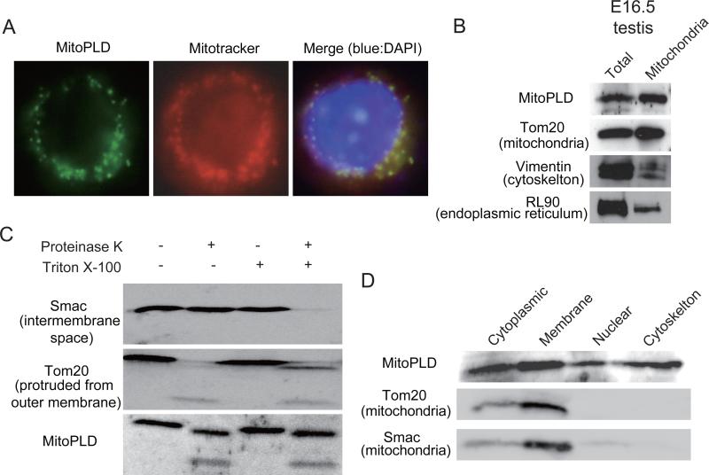
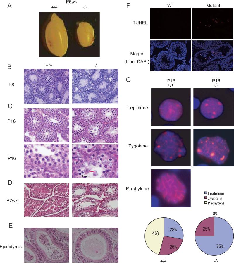
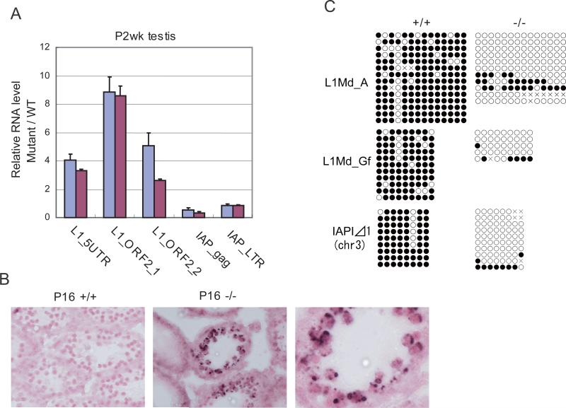
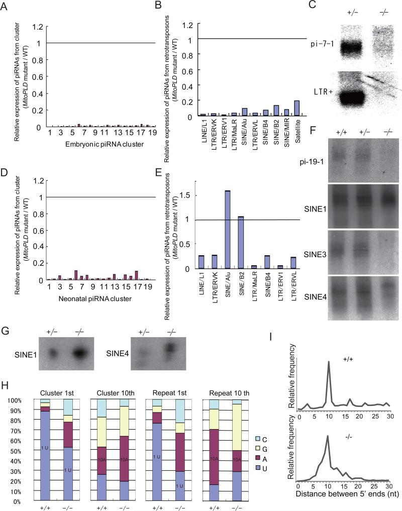
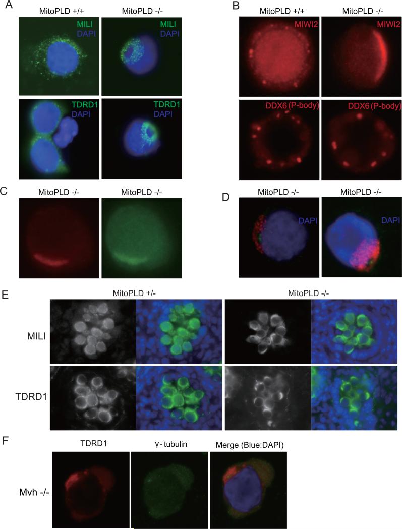
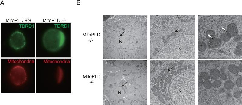
MITOPLD is a member of the phospholipase D superfamily proteins conserved among diverse species. Zucchini (Zuc), the Drosophila homolog of MITOPLD, has been implicated in primary biogenesis of Piwi-interacting RNAs (piRNAs). By contrast, MITOPLD has been shown to hydrolyze cardiolipin in the outer membrane of mitochondria to generate phosphatidic acid, which is a signaling molecule. To assess whether the mammalian MITOPLD is involved in piRNA biogenesis, we generated Mitopld mutant mice. The mice display meiotic arrest during spermatogenesis, demethylation and derepression of retrotransposons, and defects in primary piRNA biogenesis. Furthermore, in mutant germ cells, mitochondria and the components of the nuage, a perinuclear structure involved in piRNA biogenesis/function, are mislocalized to regions around the centrosome, suggesting that MITOPLD may be involved in microtubule-dependent localization of mitochondria and these proteins. Our results indicate a conserved role for MITOPLD/Zuc in the piRNA pathway and link mitochondrial membrane metabolism/signaling to small RNA biogenesis.


Figures

Figure 1
Figure 1. MitoPLD is localized to the mitochondrial outer membrane
A. Mouse MitoPLD ectopically expressed in NIH3T3 cells is localized to mitochondria. Immunostaining was performed with an antibody raised against a C-terminal peptide (left). Mitotracker labels mitochondria (center). A merged view with DAPI staining (right). B. MitoPLD is found in mitochondria of testicular cells. Mitochondria were purified by centrifugation from E16.5 testes. A total testis lysate and a mitochondrial lysate were subjected to Western blotting with antibodies against a middle portion of MitoPLD, Tom20, Vimentin and RL90 are markers for mitochondria, cytoskeleton and endoplasmic reticulum, respectively. C. MitoPLD is localized on the outer membrane of mitochondria. Mitochondria were treated with proteinase K in the presence or absence of Triton X-100. Triton X-100 disrupts the membrane structure of mitochondria. Smac is localized in the inter-membrane space and thus is digested only when Triton X-100 is present. Tom20 is protruded from the outer membrane and therefore digested by proteinase K regardless of the presence/absence of Triton-X-100. D. MitoPLD is also found in other subcellular fractions. Testicular cells were subjected to subcellular fractionation, and Western blotting was performed with antibodies against MitoPLD, Tom20 and Smac.
Figure 2
Figure 2. Meiotic defects in MitoPLD mutant testes
A. Testes from MitoPLD-/- mice were significantly smaller than those from +/+ control mice at P6wk. B. Sections from MitoPLD-/- testes show no discernable defect at P8. C. MitoPLD-/- testes show spermatocytes with abnormal morphology at P16. In higher magnification views (bottom), MitoPLD-/- testes have spermatocytes with a condensed nucleus (blue arrow) and those with a swollen nucleus (red arrow). D. MitoPLD-/- testes lack round and elongated spermatids at P7wk. E. No spermatozoa are observed in MitoPLD-/- epididymides at P8wk. F. TUNEL assays identify apoptotic cells in MitoPLD-/- testes at P6wk. G. Spermatocytes are arrested at the zygotene stage in MitoPLD-/- testes. Nuclei of spermatocytes from P16 testes were immunostained with an SYCP3 antibody (red) and counterstained with DAPI (blue). Percentage of spermatocytes at each stage of the meiotic prophase is shown at the bottom.
Figure 3
Figure 3. Retrotransposons are derepressed in MitoPLD mutant testes
A. Quantitative PCR analysis of L1Md and IAP retrotransposon RNAs in P2wk testes. Testes from two MitoPLD-/- mice were analyzed (red and blue bars). The level of Gapdh mRNA was used to normalize the data from mutants and wildtype mice. The relative RNA levels are shown. Error bars represent S. D. (n = 3). B. In situ hybridization analysis of P16 testes with a probe that detects L1Md RNA. Lump-like signals of L1Md RNA are observed around nuclei of spermatocytes only in MitoPLD-/- testes. Nuclei were stained with Nuclear Fast Red. C. DNA methylation analysis of retrotransposon sequences in P5-7 spermatogonia by bisulfite sequencing.
Figure 4
Figure 4. Defects in primary piRNA biogenesis in MitoPLD mutant testes
A. Expression of unique sequence piRNAs in MitoPLD-/- testes relative to MitoPLD+/+ testes at E16.5. The data for the piRNAs from the top 19 piRNA clusters producing the largest numbers (amounts) of piRNA in wildtype testes are shown. The positions of these clusters are shown in Supplemental Information. The piRNA counts were normalized by the miRNA counts. B. Expression of piRNAs from retrotransposons in MitoPLD-/- testes relative to MitoPLD+/+ testes at E16.5. The data for the piRNAs from the retrotransposons of top 10 piRNA sources are shown. The retrotransposons are ordered from left to right according to the number of piRNAs they produce in wildtype. The total miRNA counts were used for normalization. C. Northern blotting analysis of piRNAs in MitoPLD+/- and -/- testes at E16.5. pi-7-1 is a piRNA generated from the largest embryonic piRNA cluster on chr7. LTR+ piRNA has a sequence of IAP1 retrotransposon. D. Expression of unique sequence piRNAs in MitoPLD-/- testes relative to MitoPLD+/+ testes at P10. See (A). E. Expression of piRNAs from retrotransposons in MitoPLD-/- testes relative to MitoPLD+/+ testes at P10. The data for the top 8 sources of piRNAs in wildtype are shown. Retrotransposons are ordered from left to right according to the number of piRNAs that they produce. See (B). F. Northern blotting analysis of piRNAs in MitoPLD+/+, +/- and -/- testes at P10. pi-19-1 is a unique piRNA from a piRNA cluster. pi-SINE1, 3 and 4 have sequences of SINE B1. G. The piRNAs from SINE B1 are bound to MILI. The MILI complex was immunoprecipitated from P10 testes and the bound RNAs were probed with oligonulceotides for pi-SINE1 and pi-SINE4 in (F). RNA from five pairs of testes was loaded in each lane. H. piRNAs from MitoPLD-/- testes tend to lose the characteristics of the primary piRNA (1st U) but gain that of the secondary piRNA (complementarity between 1st base and 10th base). The nucleotide compositions of the 1st and 10th nucleotide are shown for piRNAs from clusters and repeat from P10 testes. I. The distance between the 5’ ends of complementary piRNAs. piRNAs from P10 testes that matched the consensus sequence of IAP1 retrotransposon with up to 3 mismatches were used. A peak at 10 nt in MitoPLD-/- testes suggests that the ping-pong cycle (the secondary pathway) is still active.
Figure 5
Figure 5. MitoPLD is required for nuage localization
A. Mislocalization of the pi-body components in MitoPLD-/- testes. Prospermatogonia from E16.5 testes were immunostained with MILI or TDRD1 antibodies in the presence of 0.1 % Triton X-100. B. Mislocalization of the piP-body components in MitoPLD-/- testes. Prospermatogonia from E16.5 testes were immunostained with MIWI2 or DDX6 antibodies in the absence of the detergent. C. Colocalization of MILI and MIWI2 in MitoPLD-/- testes. Prospermatogonia from E16.5 testes were double stained in the absence of detergent. The difference in MILI staining pattern between (A) and (C) (doughnut-shaped staining and crescent-like staining) results from the presence or absence of Triton X-100. D. MILI and TDRD1 are mislocalized around the centrosome in MitoPLD-/- testes. Prospermatogonia from E16.5 testes were double-stained with gamma-tubulin, a marker for the centrosome, and MILI (left) or TDRD1 (right) anitibodies. E. Mislocalization of nuage components in MitoPLD and Mvh mutants. Immunostaining of MILI and TDRD1 show crescent-like staining in MitoPLD-/- testes at E16.5. F. Immunostaining patterns of TDRD1 and gamma-tubulin are essentially the same in Mvh mutant and MitoPLD mutant prospermatogonia at E16.5.
Figure 6
Figure 6. MitoPLD is required for mitochondrial localization
A. Mitochondria are mislocalized around the centrosome and colocalize with TDRD1 in MitoPLD-/- testes. Prospermatogonia from E16.5 testes were double-stained with MitoTracker and TDRD1 antibody. B. Conglomeration of mitochondria around the nucleus and dininished pi-body formation in MitoPLD-/- prospermatogonia observed by electron microscopy. Sections from E16.5 testes were studied. Mitochondria are indicated by arrows and pi-bodies in the control testes are indicated by arrowheads. Nuclei are marked “N” in the left and middle panels.

References

    1. Anderson R, Schaible K, Heasman J, Wylie C. Expression of the homophilic adhesion molecule, Ep-CAM, in the mammalian germ line. J Reprod Fertil. 1999;116:379–384. - PubMed
    1. Aravin A, Gaidatzis D, Pfeffer S, Lagos-Quintana M, Landgraf P, Iovino N, Morris P, Brownstein MJ, Kuramochi-Miyagawa S, Nakano T, et al. A novel class of small RNAs bind to MILI protein in mouse testes. Nature. 2006;442:203–207. - PubMed
    1. Aravin AA, Hannon GJ, Brennecke J. The Piwi-piRNA pathway provides an adaptive defense in the transposon arms race. Science. 2007a;318:761–764. - PubMed
    1. Aravin AA, Sachidanandam R, Bourc'his D, Schaefer C, Pezic D, Toth KF, Bestor T, Hannon GJ. A piRNA pathway primed by individual transposons is linked to de novo DNA methylation in mice. Mol Cell. 2008;31:785–799. - PMC - PubMed
    1. Aravin AA, Sachidanandam R, Girard A, Fejes-Toth K, Hannon GJ. Developmentally regulated piRNA clusters implicate MILI in transposon control. Science. 2007b;316:744–747. - PubMed

Publication types

MeSH terms

Associated data