Clinical Trial
. 2012 Feb 1;82(2):924-32.
doi: 10.1016/j.ijrobp.2010.12.068. Epub 2011 Mar 11.

Combination of external beam radiotherapy (EBRT) with intratumoral injection of dendritic cells as neo-adjuvant treatment of high-risk soft tissue sarcoma patients

Affiliations
Clinical Trial

Combination of external beam radiotherapy (EBRT) with intratumoral injection of dendritic cells as neo-adjuvant treatment of high-risk soft tissue sarcoma patients

Steven E Finkelstein et al. Int J Radiat Oncol Biol Phys. .

Abstract

Purpose: The goal of this study was to determine the effect of combination of intratumoral administration of dendritic cells (DC) and fractionated external beam radiation (EBRT) on tumor-specific immune responses in patients with soft-tissue sarcoma (STS).

Methods and material: Seventeen patients with large (>5 cm) high-grade STS were enrolled in the study. They were treated in the neoadjuvant setting with 5,040 cGy of EBRT, split into 28 fractions and delivered 5 days per week, combined with intratumoral injection of 10(7) DCs followed by complete resection. DCs were injected on the second, third, and fourth Friday of the treatment cycle. Clinical evaluation and immunological assessments were performed.

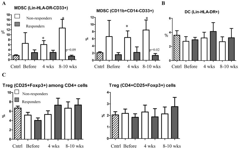
Results: The treatment was well tolerated. No patient had tumor-specific immune responses before combined EBRT/DC therapy; 9 patients (52.9%) developed tumor-specific immune responses, which lasted from 11 to 42 weeks. Twelve of 17 patients (70.6%) were progression free after 1 year. Treatment caused a dramatic accumulation of T cells in the tumor. The presence of CD4(+) T cells in the tumor positively correlated with tumor-specific immune responses that developed following combined therapy. Accumulation of myeloid-derived suppressor cells but not regulatory T cells negatively correlated with the development of tumor-specific immune responses. Experiments with (111)In labeled DCs demonstrated that these antigen presenting cells need at least 48 h to start migrating from tumor site.

Conclusions: Combination of intratumoral DC administration with EBRT was safe and resulted in induction of antitumor immune responses. This suggests that this therapy is promising and needs further testing in clinical trials design to assess clinical efficacy.


Conflict of interest statement

Conflict of interest notification

No Actual or potential conflicts of interest exist

Figures

Figure 1
Figure 1. Immune response in patients treated with EBRT and DC administration
A. IFN-γ producing cells in ELISPOT assay. Each experiment was performed in quadruplicate. Patients 12 and 10 were considered as responders, patient 7 as non-responder. B. T-cell proliferation after stimulation with 100 μg control or tumor cell lysates. Cell proliferation was evaluated by uptake of 3[H]-thymidine. Each experiment was performed in triplicate. Patients 7 and 8 were considered as responders, patient 4 as non-responder. C. MNC from indicated patients were stimulated with DCs infected with control adenovirus (Ad-c) or Ad-surv. The number of IFN-γ producing cells was evaluated in ELISPOT assay. Each experiment was performed in quadruplicate. Patients 15 and 8 were considered as responders, patient 16 as non-responder.
Figure 2
Figure 2. Immune response to survivin and T-cell infiltration of the tumors
A. Example of staining with anti-survivin antibody of tumor tissues collected prior to start of the treatment. Left panel patient with high expression of surviving (>20%), right panel patient with low expression (<20%). B. Statistical analysis of link between survivin expression in tumors and survivin-specific immune response to the treatment. Patients with less than 20% survivin positive tumor cells were considered “survivin low” and more than 20% as “survivin high”. Non-responders – patients that did not develop immune response to TAA, responders – patients who had detectable response to either TCL or survin. Tumor samples were available from only 16 patients. C. Tumor tissues collected prior to the treatment (left panel) and during the surgery after the treatment (right panel) and stained with anti-CD3 antibody. D. Tumor tissues were collected before start of the treatment and during the surgery (4–5 weeks after the treatment). For the analysis patients were split into three groups: those with robust response to vaccination (n= 5), transient response (n=3) and non-responders (n=5). The number of T-lymphocytes in tumor tissues was evaluated by immunohistochemistry and calculated per high power field (x400) after counting at least 10 randomly selected fields. For each patient group the difference between the number of cells in tumor tissues after the treatment was significantly higher than before (p<0.05). The differences between groups were not statistically significant except as shown in the figure.
Figure 3
Figure 3. Cytokines and immune suppressive myeloid and lymphoid cells
A. Proportion of MDSC in patients. Two antibody cocktails (indicated on the graphs) were used to identify MDSC. Cntrl – values in 12 healthy volunteers. * - statistically significant differences from control. P values for statistically significant differences between the groups are shown on the graph. B. Proportion of DCs in cancer patients. C. Proportion of Treg in patient.
Figure 4
Figure 4. Evaluation of DCs migration after injection to the tumor
DCs were labeled with 111In and injected intratumorally at different time prior to surgery. 24 hr group included 5 patients, 48 hr group – 4 patients, and 72 hr group – 6 patients. A. Ratio of radioactivity at the edge of the tumor to the center of the tumor. B. Ratio of radioactivity at the draining lymph nodes to the radioactivity at the center of the tumor. Background – ratio of radioactivity at distant site to the radioactivity at the center of the tumor. P values are shown on the graph

References

    1. Restifo NP, Antony PA, Finkelstein SE, et al. Assumptions of the tumor escape hypothesis. Semin Cancer Biol. 2002;12:81–86. - PMC - PubMed
    1. Friedman EJ. Immune modulation by ionizing radiation and its implications for cancer immunotherapy. Curr Pharm Des. 2002;8:1765–1780. - PubMed
    1. Overwijk WW, Theoret MR, Finkelstein SE, et al. Tumor regression and autoimmunity after reversal of a functionally tolerant state of self-reactive CD8+ T cells. J Exp Med. 2003;198:569–580. - PMC - PubMed
    1. Gattinoni L, Finkelstein SE, Klebanoff CA, et al. Removal of homeostatic cytokine sinks by lymphodepletion enhances the efficacy of adoptively transferred tumor-specific CD8+ T cells. J Exp Med. 2005;202:907–912. - PMC - PubMed
    1. Quarmby S, Kumar P, Kumar S. Radiation-induced normal tissue injury: role of adhesion molecules in leukocyte-endothelial cell interactions. Int J Cancer. 1999;82:385–395. - PubMed

Publication types

MeSH terms

LinkOut - more resources