Deletion of the p85alpha regulatory subunit of phosphoinositide 3-kinase in cone photoreceptor cells results in cone photoreceptor degeneration
- PMID: 21398281
- PMCID: PMC3109053
- DOI: 10.1167/iovs.10-7139
Deletion of the p85alpha regulatory subunit of phosphoinositide 3-kinase in cone photoreceptor cells results in cone photoreceptor degeneration
Abstract
Purpose: Downregulation of the retinal insulin/mTOR pathway in mouse models of retinitis pigmentosa is linked to cone cell death, which can be delayed by systemic administration of insulin. A classic survival kinase linking extracellular trophic/growth factors with intracellular antiapoptotic pathways is phosphoinositide 3-kinase (PI3K), which the authors have shown to protect rod photoreceptors from stress-induced cell death. The role of PI3K in cones was studied by conditional deletion of its p85α regulatory subunit.
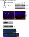
Methods: Mice expressing Cre recombinase in cones were bred to mice with a floxed pi3k gene encoding the p85α regulatory subunit of the PI3K and were back-crossed to ultimately generate offspring with cone-specific p85α knockout (cKO). Cre expression and cone-specific localization were confirmed by Western blot analysis and immunohistochemistry (IHC), respectively. Cone structural integrity was determined by IHC using peanut agglutinin and an M-opsin-specific antibody. Electroretinography (ERG) was used to assess rod and cone photoreceptor function. Retinal structure was examined by light and electron microscopy.
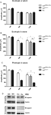
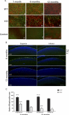
Results: An age-related cone degeneration was found in cKO mice, evidenced by a reduction in photopic ERG amplitudes and loss of cone cells. By 12 months of age, approximately 78% of cones had died, and progressive disorganization of synaptic ultrastructure was noted in surviving cone terminals in cKO retinas. Rod viability was unaffected in p85α cKO mice.
Conclusions: The present study suggests that PI3K signaling pathway is essential for cone survival in the mouse retina.
Figures


Similar articles
-
Insulin receptor signaling in cones.J Biol Chem. 2013 Jul 5;288(27):19503-15. doi: 10.1074/jbc.M113.469064. Epub 2013 May 14. J Biol Chem. 2013. PMID: 23673657 Free PMC article.
-
Phosphoinositide 3-kinase signaling in retinal rod photoreceptors.Invest Ophthalmol Vis Sci. 2011 Aug 11;52(9):6355-62. doi: 10.1167/iovs.10-7138. Invest Ophthalmol Vis Sci. 2011. PMID: 21730346 Free PMC article.
-
CNGA3 deficiency affects cone synaptic terminal structure and function and leads to secondary rod dysfunction and degeneration.Invest Ophthalmol Vis Sci. 2012 Mar 1;53(3):1117-29. doi: 10.1167/iovs.11-8168. Invest Ophthalmol Vis Sci. 2012. PMID: 22247469 Free PMC article.
-
Conditional gene knockout system in cone photoreceptors.Adv Exp Med Biol. 2006;572:173-8. doi: 10.1007/0-387-32442-9_26. Adv Exp Med Biol. 2006. PMID: 17249572 Review.
-
Thyroid Hormone Signaling and Cone Photoreceptor Viability.Adv Exp Med Biol. 2016;854:613-8. doi: 10.1007/978-3-319-17121-0_81. Adv Exp Med Biol. 2016. PMID: 26427466 Review.
Cited by
-
Beyond Genetics: The Role of Metabolism in Photoreceptor Survival, Development and Repair.Front Cell Dev Biol. 2022 May 18;10:887764. doi: 10.3389/fcell.2022.887764. eCollection 2022. Front Cell Dev Biol. 2022. PMID: 35663397 Free PMC article. Review.
-
Phosphorylated Grb14 is an endogenous inhibitor of retinal protein tyrosine phosphatase 1B, and light-dependent activation of Src phosphorylates Grb14.Mol Cell Biol. 2011 Oct;31(19):3975-87. doi: 10.1128/MCB.05659-11. Epub 2011 Jul 26. Mol Cell Biol. 2011. PMID: 21791607 Free PMC article.
-
Insulin receptor signaling in cones.J Biol Chem. 2013 Jul 5;288(27):19503-15. doi: 10.1074/jbc.M113.469064. Epub 2013 May 14. J Biol Chem. 2013. PMID: 23673657 Free PMC article.
-
The p110α isoform of phosphoinositide 3-kinase is essential for cone photoreceptor survival.Biochimie. 2015 May;112:35-40. doi: 10.1016/j.biochi.2015.02.018. Epub 2015 Mar 3. Biochimie. 2015. PMID: 25742742 Free PMC article.
-
Metabolic and redox signaling in the retina.Cell Mol Life Sci. 2017 Oct;74(20):3649-3665. doi: 10.1007/s00018-016-2318-7. Epub 2016 Aug 20. Cell Mol Life Sci. 2017. PMID: 27543457 Free PMC article. Review.
References
-
- Cantley LC. The phosphoinositide 3-kinase pathway. Science. 2002;296:1655–1657 - PubMed
-
- Dudek H, Datta SR, Franke TF, et al. Regulation of neuronal survival by the serine-threonine protein kinase Akt. Science. 1997;275:661–665 - PubMed
-
- Fukami K, Endo T, Imamura M, Takenawa T. alpha-Actinin and vinculin are PIP2-binding proteins involved in signaling by tyrosine kinase. J Biol Chem. 1994;269:1518–1522 - PubMed
-
- Fukami K, Sawada N, Endo T, Takenawa T. Identification of a phosphatidylinositol 4,5-bisphosphate-binding site in chicken skeletal muscle alpha-actinin. J Biol Chem. 1996;271:2646–2650 - PubMed
-
- Guo X, Ghalayini AJ, Chen H, Anderson RE. Phosphatidylinositol 3-kinase in bovine photoreceptor rod outer segments. Invest Ophthalmol Vis Sci. 1997;38:1873–1882 - PubMed
Publication types
MeSH terms
Substances
Grants and funding
LinkOut - more resources
Full Text Sources
Molecular Biology Databases
Miscellaneous

