. 2011 May;300(5):L730-9.
doi: 10.1152/ajplung.00308.2010. Epub 2011 Mar 11.

C-peptide, a novel inhibitor of lung inflammation following hemorrhagic shock

Affiliations

C-peptide, a novel inhibitor of lung inflammation following hemorrhagic shock

Ranjit S Chima et al. Am J Physiol Lung Cell Mol Physiol. 2011 May.

Abstract

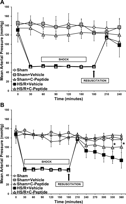
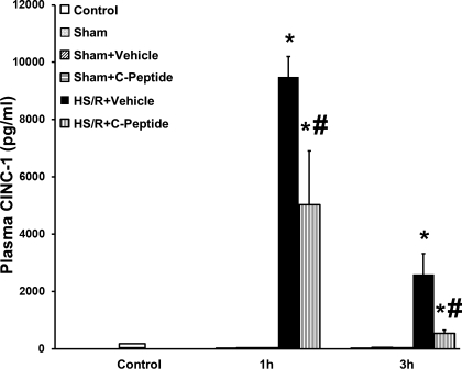
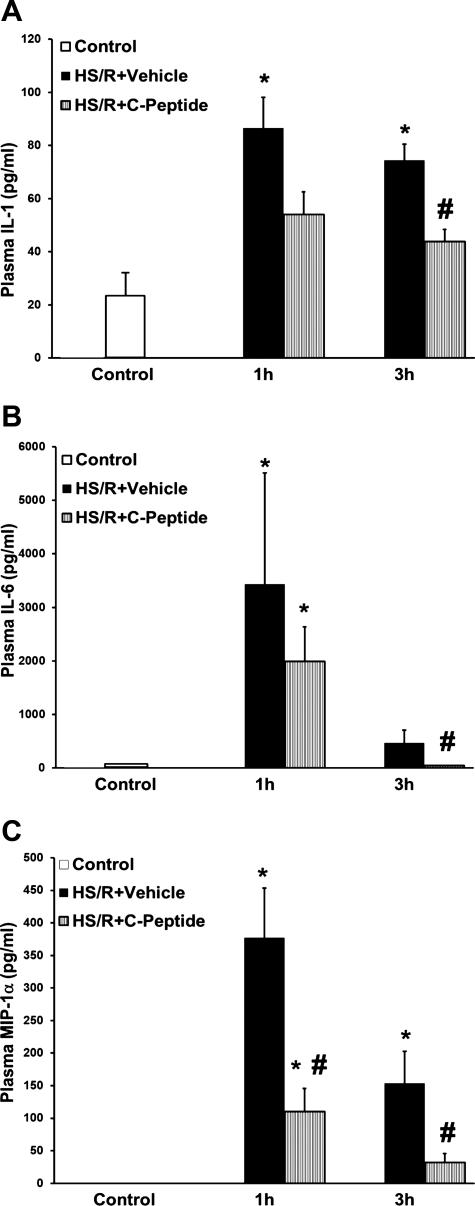
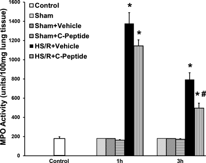
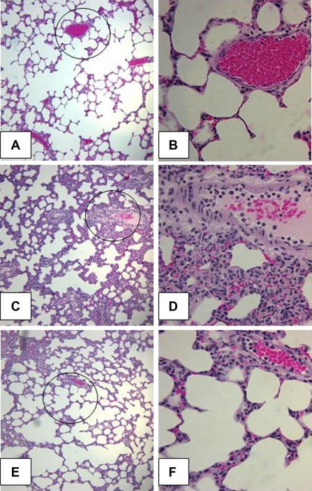
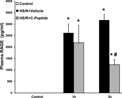
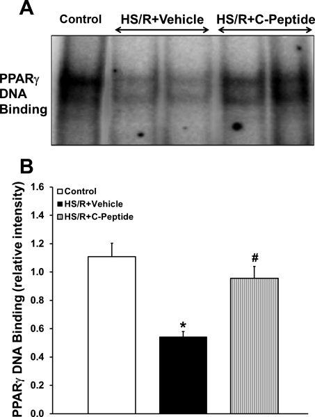
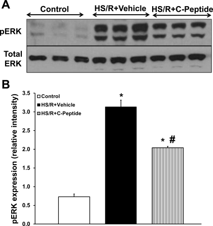
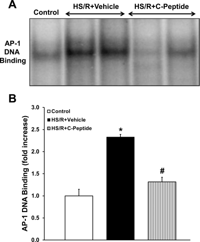
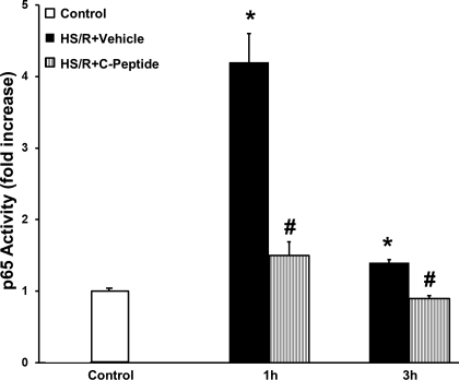
C-peptide is a 31-amino acid peptide cleaved from proinsulin during insulin synthesis. Initially thought to be inert, C-peptide may modulate the inflammatory response in the setting of endotoxemia and ischemia reperfusion. However, the spectrum of its biological effects is unclear. We hypothesized that exogenous administration of C-peptide would modulate pro- and anti-inflammatory signaling pathways and thereby attenuate lung inflammation in an in vivo model of hemorrhagic shock. Hemorrhagic shock was induced in male Wistar rats (aged 3-4 mo) by withdrawing blood to a mean arterial pressure of 50 mmHg. At 3 h after hemorrhage, rats were rapidly resuscitated by returning their shed blood. At the time of resuscitation and every hour thereafter, animals received C-peptide (280 nmol/kg) or vehicle parenterally. Animals were euthanized at 1 and 3 h after resuscitation. C-peptide administration at resuscitation following hemorrhagic shock ameliorated hypotension and blunted the systemic inflammatory response by reducing plasma levels of IL-1, IL-6, macrophage inflammatory protein-1α, and cytokine-induced neutrophil chemoattractant-1. This was associated with a reduction in lung neutrophil infiltration and plasma levels of receptor for advanced glycation end products. Mechanistically, C-peptide treatment was associated with reduced expression of proinflammatory transcription factors activator protein-1 and NF-κB and activation of the anti-inflammatory transcription factor peroxisome proliferator-activated receptor-γ. Our data suggest that C-peptide ameliorates the inflammatory response and lung inflammation following hemorrhagic shock. These effects may be modulated by altering the balance between pro- and anti-inflammatory signaling in the lung.


Figures

Fig. 1.
Fig. 1.
Effect of in vivo treatment with vehicle or C-peptide on mean arterial pressure (MAP). A: time course depiction of MAP up to 1 h following resuscitation. B: time course depiction of MAP up to 3 h following resuscitation. Each data point represents mean ± SD of 3 rats in sham groups and 5 rats in hemorrhagic shock and resuscitation (HS/R) groups. *P < 0.05 vs. HS/R+Vehicle group.
Fig. 2.
Fig. 2.
Effect of in vivo treatment with vehicle or C-peptide on plasma cytokine-induced neutrophil chemoattractant 1 (CINC-1) levels at 1 and 3 h following HS/R. Each data point represents mean ± SE of 3 rats in sham groups, 5 rats in control and HS/R groups. *P < 0.05 vs. Control group; #P < 0.05 vs. HS/R+Vehicle group.
Fig. 3.
Fig. 3.
Effect of in vivo treatment with vehicle or C-peptide on plasma levels of IL-1 (A), IL-6 (B), and macrophage inflammatory protein-1 α (MIP-1α; C) at 1 and 3 h following HS/R. Each data point represents mean ± SE of 5 rats. *P < 0.05 vs. Control group; #P < 0.05 vs. HS/R+Vehicle group.
Fig. 4.
Fig. 4.
Effect of in vivo treatment with vehicle or C-peptide on lung MPO activity at 1 and 3 h following HS/R. Each data point represents mean ± SE of 3 rats in sham groups, 5 rats in basal and HS/R groups. *P < 0.05 vs. Control group; #P < 0.05 vs. HS/R+Vehicle group.
Fig. 5.
Fig. 5.
Effect of in vivo treatment with vehicle or C-peptide on lung histology at 3 h after HS/R. A: representative photomicrograph of histology showing lung architecture from a rat in the Control group (magnification ×100). B: magnified section of prior image (indicated by circle) to show alveolar architecture and blood vessel from a rat in the Control group (magnification ×400). C: representative photomicrograph of histology showing lung architecture from a rat in the HS/R+Vehicle group (magnification ×100). D: magnified section of prior image (indicated by circle) demonstrating interstitial edema and inflammatory cell infiltration from a rat in the HS/R+Vehicle group (magnification ×400). E: representative photomicrograph of histology showing lung architecture from a rat in the HS/R+C-peptide group (magnification ×100). F: magnified section of prior image (indicated by circle) demonstrating a reduction in interstitial edema and inflammatory cell infiltration from a rat in the HS/R+C-peptide group (magnification ×400).
Fig. 6.
Fig. 6.
Effect of in vivo treatment with vehicle or C-peptide on plasma receptor for advanced glycation end products (RAGE) level at 1 and 3 h following HS/R. Each data point represents mean ± SE of 5 rats. *P < 0.05 vs. Control group; #P < 0.05 vs. HS/R+Vehicle group.
Fig. 7.
Fig. 7.
Effect of in vivo treatment with vehicle or C-peptide on PPAR-γ DNA binding in lung at 3 h following HS/R. A: representative autoradiograph of EMSA for PPAR-γ. B: image analysis of PPAR-γ DNA binding determined by densitometry. Each data point represents mean ± SE of 5 rats from 2 separate experiments. *P < 0.05 vs. Control group; #P < 0.05 vs. HS/R+Vehicle group.
Fig. 8.
Fig. 8.
Effect of in vivo treatment with vehicle or C-peptide on phosphorylated form of ERK (pERK) in lung cytosol extracts at 3 h following HS/R. A: representative Western blot of phosphorylated form of ERK and total ERK. B: image analysis of nuclear content of phosphorylated form of ERK as determined by densitometry. Each data point represents mean ± SE of 5 rats from 2 separate experiments. *P < 0.05 vs. Control group; #P < 0.05 vs. HS/R+Vehicle group.
Fig. 9.
Fig. 9.
Effect of in vivo treatment with vehicle or C-peptide on activator protein-1 (AP-1) DNA binding in lung at 3 h following HS/R. A: representative autoradiograph of EMSA for AP-1. B: image analysis of AP-1 DNA binding determined by densitometry. Fold increase was calculated vs. control value, which was set at 1.0. Each data point represents mean ± SE of 5 rats from 2 separate experiments. *P < 0.05 vs. Control group; #P < 0.05 vs. HS/R+Vehicle group.
Fig. 10.
Fig. 10.
Effect of in vivo treatment with vehicle or C-peptide on activation of p65 subunit of NF-κB in lung nuclear extracts as determined by transcription factor assay at 1 and 3 h following HS/R. Fold increase was calculated vs. control value which was set at 1.0. Each data point represents mean ± SE of 5 rats. *P < 0.05 vs. Control group; #P < 0.05 vs. HS/R+Vehicle group.

Similar articles

Cited by

References

    1. Abraham E. Neutrophils and acute lung injury. Crit Care Med 31: S195–S199, 2003 - PubMed
    1. Abraham E. NF-kappaB activation. Crit Care Med 28: N100–N104, 2000 - PubMed
    1. Adams M, Reginato MJ, Shao D, Lazar MA, Chatterjee VK. Transcriptional activation by peroxisome proliferator-activated receptor gamma is inhibited by phosphorylation at a consensus mitogen-activated protein kinase site. J Biol Chem 272: 5128–5132, 1997 - PubMed
    1. Al-Rasheed NM, Chana RS, Baines RJ, Willars GB, Brunskill NJ. Ligand-independent activation of peroxisome proliferator-activated receptor-gamma by insulin and C-peptide in kidney proximal tubular cells: dependent on phosphatidylinositol 3-kinase activity. J Biol Chem 279: 49747–49754, 2004 - PubMed
    1. Al-Rasheed NM, Meakin F, Royal EL, Lewington AJ, Brown J, Willars GB, Brunskill NJ. Potent activation of multiple signalling pathways by C-peptide in opossum kidney proximal tubular cells. Diabetologia 47: 987–997, 2004 - PubMed

Publication types

MeSH terms