. 2011 May 6;286(18):16039-51.
doi: 10.1074/jbc.M110.204057. Epub 2011 Mar 11.

Mechanistic control of carcinoembryonic antigen-related cell adhesion molecule-1 (CEACAM1) splice isoforms by the heterogeneous nuclear ribonuclear proteins hnRNP L, hnRNP A1, and hnRNP M

Affiliations

Mechanistic control of carcinoembryonic antigen-related cell adhesion molecule-1 (CEACAM1) splice isoforms by the heterogeneous nuclear ribonuclear proteins hnRNP L, hnRNP A1, and hnRNP M

Kenneth J Dery et al. J Biol Chem. .

Abstract

Carcinoembryonic antigen-related cell adhesion molecule-1 (CEACAM1) is expressed in a variety of cell types and is implicated in carcinogenesis. Alternative splicing of CEACAM1 pre-mRNA generates two cytoplasmic domain splice variants characterized by the inclusion (L-isoform) or exclusion (S-isoform) of exon 7. Here we show that the alternative splicing of CEACAM1 pre-mRNA is regulated by novel cis elements residing in exon 7. We report the presence of three exon regulatory elements that lead to the inclusion or exclusion of exon 7 CEACAM1 mRNA in ZR75 breast cancer cells. Heterologous splicing reporter assays demonstrated that the maintenance of authentic alternative splicing mechanisms were independent of the CEACAM1 intron sequence context. We show that forced expression of these exon regulatory elements could alter CEACAM1 splicing in HEK-293 cells. Using RNA affinity chromatography, three members of the heterogeneous nuclear ribonucleoprotein family (hnRNP L, hnRNP A1, and hnRNP M) were identified. RNA immunoprecipitation of hnRNP L and hnRNP A1 revealed a binding motif located central and 3' to exon 7, respectively. Depletion of hnRNP A1 or L by RNAi in HEK-293 cells promoted exon 7 inclusion, whereas overexpression led to exclusion of the variable exon. By contrast, overexpression of hnRNP M showed exon 7 inclusion and production of CEACAM1-L mRNA. Finally, stress-induced cytoplasmic accumulation of hnRNP A1 in MDA-MB-468 cells dynamically alters the CEACAM1-S:CEACAM1:L ratio in favor of the l-isoform. Thus, we have elucidated the molecular factors that control the mechanism of splice-site recognition in the alternative splicing regulation of CEACAM1.


Figures
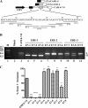
FIGURE 1.
FIGURE 1.
Identification of cis-acting regulatory elements in exon 7. A, shown is a schematic of scanning mutagenesis across exon 7 in CAM 6-7-8 reporter and derivative mini-genes. The splicing of CAM 6-7-8 yields CEACAM1 long and short cytoplasmic domain splice variants, CEACAM1-S and CEACAM1-L. The black rectangle represents exon 6, the gray rectangle represents exon 7, and the white rectangle represents exon 8 of CEACAM1. The black arrow represents the CMV promoter. The 10-nt block mutations are indicated: E7-1, bases 1–10; E7-2, bases 6–15; E7-3, bases 11–20; E7-4, bases 16–25; E7-5, bases 21–30; E7-6, bases 26–35; E7–7, bases 31–40; E7-8, bases 36–45; E7-9, bases 41–50. B and C, RT-PCR analysis of RNAs derived from ZR75 cells transiently transfected with indicated mini-genes or no plasmid mock control is shown. ERE-1, ERE-2, and ERE-3 refer to ERE 1, 2, and 3, respectively. M refers to a 100-bp DNA ladder (New England Biolabs). L and S refer to CEACAM1-L and CEACAM1-S. The data shown are representative of three independent experiments. The mean ± S.E. is shown for percent exon 7 inclusion calculated as the % CEACAM1-L/(CEACAM1-L + CEACAM1-S) mRNA. *, p < 0.05; **, p < 0.01; ***, p < 0.001; ****, p < 0.0001 versus CAM 6-7-8 control.
FIGURE 2.
FIGURE 2.
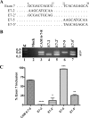
Identification of an ESE and ESS encoded within ERE-1 and ERE-2 of exon 7. A, nucleotide comparison of target ESE and ESS sequences is shown. E7-2 ′ and E7-5′ contain the wild-type sequence except where indicated with substitutions. RNAs were of equal length in relation to exon 7 but for simplicity only those nucleotides under study are depicted. B and C, RT-PCR analysis and quantitation of RNAs derived from ZR75 cells were as described in the Fig. 1 caption. *, p < 0.05; **, p < 0.01; ***, p < 0.001; ****, p < 0.0001 versus CAM 6-7-8 control.
FIGURE 3.
FIGURE 3.
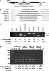
Exon 7 EREs can function in a heterologous context. A, shown is a diagram of middle exon replacement strategy in RG6 mini-gene. Black rectangles represent exons of RG6, black lines represent chicken cardiac troponin T (cTNT) introns, and the black arrow represents the CMV promoter and restriction sites unique to the middle exon (B is BamHI, and E is EcoRI). Flanking intronic sequences to the variable exon are shown in italics. The locations of vector-specific primers flanking the first and last exons are indicated with small black arrows. B, shown is a nucleotide comparison between constructs to show purine content and similarity in variable exon length. Bold nucleotides represent unique sequence inserted into middle exon of RG6. IgM-Sc and IgM-E are scrambled control (IgM-SC) and enhancer sequences from IgM substrate, respectively. CAM1-I6D5 contains an intronic splicing silencer from intron 6 of CEACAM1. C, RT-PCR after transfection in ZR75 cells with the indicated mini-genes is shown. D, titration by ERE-containing constructs alters CEACAM1 alternative splicing. RT-PCR after transfection in HEK-293 cells of CAM 6-7-8 mini-gene influenced by overexpression of indicated constructs is shown. M is Benchtop PCR marker (Promega). The mean ± S.E. for at least n = 3 is shown for percent exon 7 inclusion. *, p < 0.05; **, p < 0.01; ***, p < 0.001 versus RG6 or CAM 6-7-8 control.
FIGURE 4.
FIGURE 4.
A spliceosome regulatory complex specifically assembles on exon 7. A, unlabeled competitor RNA can titrate an exon 7-associated nuclear complex. Left panel, 32P-labeled exon 7 RNA (0.28 pmol) was incubated in the presence (+) (lanes 2–8) or absence (−) (lane 1) of HeLa nuclear extract (NE) under splicing conditions and resolved on a native gel. Unlabeled competitor RNA was also added to the reactions as indicated. Right panel, an overexposed membrane (lanes 6–8) of the left panel is shown. B, UV light-induced cross-linking to exon 7 reveals nuclear and cytoplasmic proteins. S100 extract (containing cytoplasmic proteins) cross-linking reactions were loaded in lanes 3 and 5. The HeLa extract (containing nuclear proteins)-cross-linking reactions were loaded in lanes 1, 2, 4, and 6. Mock-treated reactions that did not include UV cross-linking but did include proteinase K (PK) were loaded in lanes 1 and 2, respectively. C, unlabeled competitor RNA titrates cross-linked proteins associated with exon 7. HeLa nuclear extract was incubated with no unlabeled RNA competitor (lane 2), NS competitor (lanes 3 and 4) or exon 7 competitor RNA (lanes 5 and 6) as indicated. A mock-treated reaction that did not include UV cross-linking was loaded as a control (lane 1). The protein size markers (kDa) are indicated to the left, and arrows denote sites of cross-linking bands of interest.
FIGURE 5.
FIGURE 5.
RNA affinity purification of hnRNPs associated with exon 7. A, RNAs produced by transcription were used to link to adipic acid beads followed by incubation with 250 μg of HeLa nuclear extract under splicing conditions followed by SDS-PAGE electrophoresis and mass spectrometry or analyses by silver staining as shown. B, a Western blot of samples used for SDS-PAGE gel from panel A with specified antibodies is shown.
FIGURE 6.
FIGURE 6.
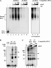
Binding and gene dosage analyses of hnRNP L in exon 7. A, 32P-labeled RNA was prepared using the DNA templates described in Fig. 1. Nuclear extract was incubated under splicing conditions with 0.28 pmol of uniformly labeled RNA as indicated, treated with UV light, digested with RNase A, and then either directly resolved on a 12.5% SDS-PAGE gel (odd lanes) or immunoprecipitated with anti-hnRNP L (even lanes). B, depletion and overexpression of hnRNP L results in the increased production of CEACAM1-L and CEACAM1-S, respectively. Immunoblot analyses of HEK-293 cells using anti-hnRNP L is shown. C, RT-PCR analysis of mini-genes from B (sh-Luc is directed to luciferase, sh-L is directed to hnRNP L, and FLAG-L is overexpression) is shown. The mean ± S.E. for at least n = 3 is shown. The arrow denotes cross-linking and/or immunoprecipitated hnRNP L. **, p < 0.01 versus sh-Luc control.
FIGURE 7.
FIGURE 7.
Binding and gene dosage analyses of hnRNP A1 in exon 7. A, unlabeled competitor RNA titrates hnRNP A1 association with exon 7 from nuclear extracts. Nuclear extract was incubated under splicing conditions with 0.28 pmol of uniformly labeled 32P-labeled NS RNA, 32P-labeled exon 7 RNA, and unlabeled competitor as indicated, treated with UV light, digested with RNase A, and then either directly resolved on a 12.5% SDS-PAGE gel (lanes 1 and 4) or immunoprecipitated with anti-hnRNP A1 (lanes 2, 3, 5, 6, and 7). B, hybrid NS and exon 7 RNA probes were used to narrow the hnRNP A1 binding site to the 3′ terminal end of exon 7. The schematic above the figure shows the NS RNA (white box), E7 RNA (black box), and the 20-nt ERE-1, ERE-2, and ERE-3 RNAs (black box) fused to NS RNA to make a 53-nt RNA molecule. C, depletion and overexpression of hnRNP A1 results in the increased production of CEACAM1-L and CEACAM1-S, respectively. Immunoblot analyses of HEK-293 cells using anti-hnRNP A1 is shown. D, RT-PCR analysis of mini-genes from C (sh-GFP is directed to GFP, sh-A1B is directed to hnRNP A1, and T7-A1 is over-expression) is shown. The mean ± S.E. for at least n = 3 is shown. Arrows denote cross-linking and/or immunoprecipitated hnRNP A1. **, p < 0.01 versus vector control.
FIGURE 8.
FIGURE 8.
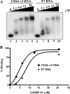
HnRNP A1-exon 7 electromobility shift assay. A, 20 fmol of CD44 variant exon v.5 RNA or 20 fmol exon 7 RNA incubated in the presence of varying amounts of His-hnRNP A1 protein (0.113–0.9 μg) and 1.0 μg of yeast tRNA to quench nonspecific interactions is shown. Reactions were electrophoresed on a 5% acrylamide gel and set to an autoradiograph. B, the apparent KD determination where the RNA-protein complexes (protein concentration for which 50% of the input is shifted to an RNP complex) were calculated as a function of increasing hnRNP A1 concentrations. The Kd values were calculated using Prism Software Version 4 (GraphPad Software). The mean ± S.E. for n = 3 was too small to be visualized with p value < 0.05 versus control.
FIGURE 9.
FIGURE 9.
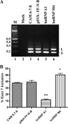
HnRNP M activates alternative splicing of CEACAM1-L mRNA. A and B, shown is transient co-transfection of CAM 6-7-8 (lane 3) and plasmid-encoded hnRNP M (lane 6) compared with hnRNP A1 (lane 5) or vector (lane 4) alone in HEK-293 cells. RT-PCR analysis and quantitation of RNAs were exactly as described in caption to Fig. 1. *, p < 0.05; **, p < 0.01 versus CAM 6-7-8.
FIGURE 10.
FIGURE 10.
Cytoplasmic accumulation of hnRNP A1 in stress-induced breast cancer cells dynamically shifts the production of CEACAM1 from the S-isoform to the L-isoform. A, HnRNP A1 localizes to stress granules in the cytoplasm. MDA-MB-468 cells cultured on an 8-well chamber slide were left untreated (−OSM) or treated (+OSM) with 0.6 m sorbitol for 2 h at 37 °C. The cells were fixed and immunostained with anti-hnRNP A1 or anti-hnRNP U antibodies to detect the endogenous proteins. BF, bright field. B and C, RT-PCR analysis and quantitation of RNA derived from cells from panel A were exactly as described in caption to Fig. 1. M is Benchtop PCR marker. *, p < 0.05 versus 0 m control.

Similar articles

Cited by

References

    1. Kuespert K., Pils S., Hauck C. R. (2006) Curr. Opin. Cell Biol. 18, 565–571 - PMC - PubMed
    1. Nittka S., Günther J., Ebisch C., Erbersdobler A., Neumaier M. (2004) Oncogene 23, 9306–9313 - PubMed
    1. Hsieh J. T., Luo W., Song W., Wang Y., Kleinerman D. I., Van N. T., Lin S. H. (1995) Cancer Res. 55, 190–197 - PubMed
    1. Riethdorf L., Lisboa B. W., Henkel U., Naumann M., Wagener C., Löning T. (1997) J. Histochem. Cytochem. 45, 957–963 - PubMed
    1. Gray-Owen S. D., Blumberg R. S. (2006) Nat. Rev. Immunol. 6, 433–446 - PubMed

Publication types

MeSH terms

Substances