. 2011 Apr 26;108(17):6811-6.
doi: 10.1073/pnas.1015248108. Epub 2011 Mar 11.

Academic cross-fertilization by public screening yields a remarkable class of protein phosphatase methylesterase-1 inhibitors

Affiliations

Academic cross-fertilization by public screening yields a remarkable class of protein phosphatase methylesterase-1 inhibitors

Daniel A Bachovchin et al. Proc Natl Acad Sci U S A. .

Abstract

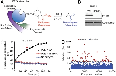
National Institutes of Health (NIH)-sponsored screening centers provide academic researchers with a special opportunity to pursue small-molecule probes for protein targets that are outside the current interest of, or beyond the standard technologies employed by, the pharmaceutical industry. Here, we describe the outcome of an inhibitor screen for one such target, the enzyme protein phosphatase methylesterase-1 (PME-1), which regulates the methylesterification state of protein phosphatase 2A (PP2A) and is implicated in cancer and neurodegeneration. Inhibitors of PME-1 have not yet been described, which we attribute, at least in part, to a dearth of substrate assays compatible with high-throughput screening. We show that PME-1 is assayable by fluorescence polarization-activity-based protein profiling (fluopol-ABPP) and use this platform to screen the 300,000+ member NIH small-molecule library. This screen identified an unusual class of compounds, the aza-β-lactams (ABLs), as potent (IC(50) values of approximately 10 nM), covalent PME-1 inhibitors. Interestingly, ABLs did not derive from a commercial vendor but rather an academic contribution to the public library. We show using competitive-ABPP that ABLs are exquisitely selective for PME-1 in living cells and mice, where enzyme inactivation leads to substantial reductions in demethylated PP2A. In summary, we have combined advanced synthetic and chemoproteomic methods to discover a class of ABL inhibitors that can be used to selectively perturb PME-1 activity in diverse biological systems. More generally, these results illustrate how public screening centers can serve as hubs to create spontaneous collaborative opportunities between synthetic chemistry and chemical biology labs interested in creating first-in-class pharmacological probes for challenging protein targets.


Conflict of interest statement

The authors declare no conflict of interest.

Figures

Fig. 1.
Fig. 1.
A fluopol-ABPP assay for PME-1. (A) Schematic representation of the reversible C-terminal methylation of PP2A introduced by LCMT1 and removed by PME-1. (B) FP-Rh (75 nM) labels purified wild-type PME-1 (1 μM) but not the catalytically dead S156A PME-1 mutant (1 μM). Top panel: Fluorescent gel image shown in gray scale. (C) Time course for fluopol signal generated by reactions of wild-type PME-1 (1 μM) with FP-Rh (75 nM). No signal increase is observed in the absence of enzyme or with the S156A PME-1 mutant. The indicated 45-min time point (Z = 0.77), prior to reaction completion, was selected for HTS. Data are presented as mean values ± SD. (D) Screening data for a representative 15,000 compounds of the NIH small-molecule library. Compounds that reduced the FP-Rh fluopol signal by > 26.13% were designated as hits for PME-1 (red squares).
Fig. 2.
Fig. 2.
Discovery of selective ABL inhibitors of PME-1. (A) Four ABLs (ABL127, ABL103, ABL105, and ABL107) identified as active in the MLPCN screen. (B) Chemistry space coordinates for compounds (colored red in high occupancy cells; colored blue in low occupancy cells) that were screened against PME-1. The 26 ABLs (shown as enlarged squares; all other compounds are shown as small circles) are located in a sparsely populated region of chemical space. This optimized 6-dimensional BCUTs (31) visualization of chemistry space for the compound library was generated using the standard 3D hydrogen suppressed descriptors with Diverse Solutions (Diverse Solutions, Tripos), with coverage illustrated by compression into an optimized two-dimensional BCUTs space as described in more detail previously (32). (C) Evaluation of compounds by gel-based competitive ABPP with FP-Rh (2 μM) in the soluble proteome (1 mg/mL protein) of MDA-MB-231 cells. (D) Complete PME-1 IC50 curves for ABL127 (4.2 nM; 95% confidence limits 2.3–7.5 nM) and ent-ABL127 (450 nM; 95% confidence limits 240–850 nM) in the soluble proteome of MDA-MB-231 cells (for IC50 curves of ABL103, ABL105, and ABL107, see Fig. S2). Data are presented as mean values ± SEM; n = 3/group. (E) Pretreatment of HEK 293T proteomes with ABL127 (500 nM, 30 min) before addition of purified PME-1 (500 nM, 1 h) blocks PP2A demethylation as determined by Western blotting with antibodies that recognize specific methylation states of PP2A.
Fig. 3.
Fig. 3.
ABLs are covalent inhibitors of PME-1. (A) Proposed mechanism for covalent PME-1 inhibition by ABL127. (B) PME-1 (500 nM) was incubated with DMSO or ABL127 (5 μM), and each reaction was split into two fractions. One fraction was directly labeled with FP-Rh (left panels), and the other was subjected to gel-filtration to remove free inhibitor and then reacted with FP-Rh (right panels) to determine the reversibility of inhibition. (C) MS1 traces for the PME-1 active site peptide not adducted (top panel) or adducted (bottom panel) to ABL127. See SI Materials and Methods for more information on this experiment. Traces were obtained from tryptic digests of purified, recombinant PME-1 (10 μM) treated with ABL127 (50 μM, red trace) or DMSO (black trace).
Fig. 4.
Fig. 4.
ABL127 selectively inactivates PME-1 and decreases the demethylated form of PP2A in cells. (A) Gel-based competitive ABPP of the soluble proteomes from MDA-MB-231 cells treated with ABL127 (0.61–10,000 nM; 1 h) reveals selective blockade of PME-1 with (B) an IC50 value of 11.1 nM. (C) Isotopically “light” and “heavy” MDA-MB-231 cells were treated with DMSO or ABL127 (100 nM), respectively, for 1 h. Proteomes were combined in a 1∶1 total protein ratio (0.5 mg each), analyzed by ABPP-MudPIT, and serine hydrolase activities were quantified by comparing intensities of light and heavy peptide peaks (see Fig. S4 for additional SILAC ABPP-MudPIT analyses). Data are presented as mean values ± SEM for all quantifiable peptides from each serine hydrolase. (D and E) MDA-MB-231 and HEK 293T cells treated with ABL127 (500 nM, 1 h) exhibit significant reductions in demethylated PP2A. (F) Stable overexpression of PME-1 in HEK 293T cells compared to control cells overexpressing GFP. PME-1 is completely inhibited by ABL127 (500 nM, 1 h) in both cell lines. (G and H) Cells overexpressing PME-1 show decreased PP2A methylation, which is reversed by addition of ABL127 (500 nM, 1 h). (I) Time-course for PME-1 inhibition by ABL127 (500 nM) in PME-1-overexpressing HEK 293T cells. For E and I: *p < 0.05, **p < 0.01 for DMSO-treated versus ABL127-treated cells. #p < 0.05, ##p < 0.01 for cells overexpressing GFP versus PME-1. Data are presented as mean values ± SEM; n = 3/group.
Fig. 5.
Fig. 5.
A clickable ABL reveals proteome-wide selectivity for PME-1. (A) Structure of the ABL alkyne probe ABL112. (B) ABL112 inactivates PME-1 (IC50 = 13.8 nM; 95% confidence limits 6.5–29 nM) in the MDA-MB-231 soluble proteome (1 mg/mL protein) as determined by gel-based competitive ABPP (see Fig. S5). (C) MDA-MB-231 cells were incubated with DMSO or ABL127 (100 nM, 30 min) followed by ABL112 (10–200 nM, 2 h). Soluble cell proteomes (0.5 mg/mL) were then subjected to a standard “click” reaction using RhN3 (30) and ABL112-labeled proteins were visualized by in-gel fluorescence scanning. PME-1 was the only protein labeled by ABL112 and competed by ABL127. ABL112 labeled an additional 80 kDa protein that was not competed by ABL127, suggesting it may be a target for ABL112, but not ABL127 (see Fig. S5). (D) Mouse brain soluble lysates (1 mg/mL) were incubated (30 min) with DMSO or ABL127 (100 nM). ABL112 (10 nM) was then added for an additional 30 minutes before analysis by click chemistry-based ABPP.
Fig. 6.
Fig. 6.
ABL127 selectively inhibits PME-1 in vivo. (A) Evaluation of mouse brain soluble proteome from mice treated with vehicle or ABL127 (50 mg kg-1, i.p., 2 h) by gel-based ABPP. In this analysis, proteomes were also treated ex vivo with DMSO or ABL127 (2 μM) before FP-Rh labeling to confirm complete PME-1 inhibition. (B) ABPP-MudPIT analysis of serine hydrolases from brain proteomes of animals treated with vehicle or ABL127. These data confirm selective inactivation of PME-1 in vivo. (C and D) Mice treated with ABL127 exhibited reductions in demethylated PP2A compared with vehicle-treated mice. *p < 0.01, **p < 0.001 for DMSO versus ABL127 groups. Data are presented as mean values ± SEM; n = 3/group.

References

    1. Manning G, Whyte DB, Martinez R, Hunter T, Sudarsanam S. The protein kinase complement of the human genome. Science. 2002;298:1912–1934. - PubMed
    1. Shi Y. Serine/threonine phosphatases: Mechanism through structure. Cell. 2009;139:468–484. - PubMed
    1. Oliver CJ, Shenolikar S. Physiologic importance of protein phosphatase inhibitors. Front Biosci. 1998;3:D961–972. - PubMed
    1. Janssens V, Goris J. Protein phosphatase 2A: A highly regulated family of serine/threonine phosphatases implicated in cell growth and signalling. Biochem J. 2001;353:417–439. - PMC - PubMed
    1. Kalhor HR, Luk K, Ramos A, Zobel-Thropp P, Clarke S. Protein phosphatase methyltransferase 1 (Ppm1p) is the sole activity responsible for modification of the major forms of protein phosphatase 2A in yeast. Arch Biochem Biophys. 2001;395:239–245. - PubMed

Publication types

MeSH terms

Substances