. 2011 Jun;300(6):H2230-7.
doi: 10.1152/ajpheart.01226.2010. Epub 2011 Mar 11.

Arrhythmogenic substrate in hearts of rats with monocrotaline-induced pulmonary hypertension and right ventricular hypertrophy

Affiliations

Arrhythmogenic substrate in hearts of rats with monocrotaline-induced pulmonary hypertension and right ventricular hypertrophy

David Benoist et al. Am J Physiol Heart Circ Physiol. 2011 Jun.

Abstract

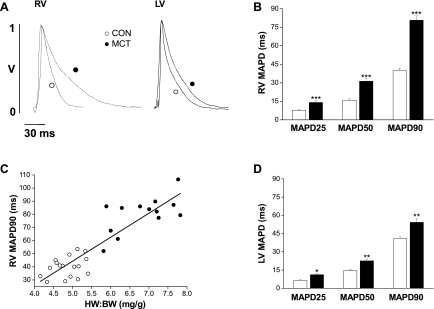
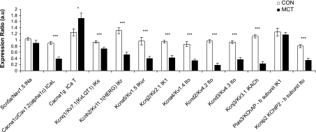
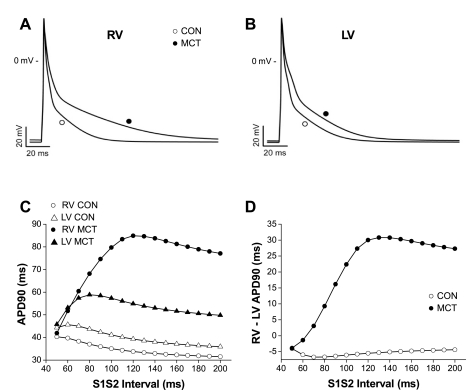
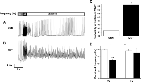
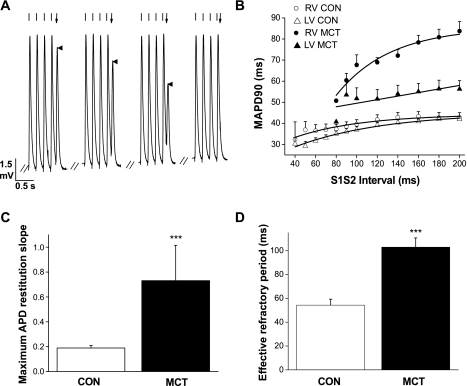
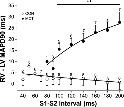
Mechanisms associated with right ventricular (RV) hypertension and arrhythmias are less understood than those in the left ventricle (LV). The aim of our study was to investigate whether and by what mechanisms a proarrhythmic substrate exists in a rat model of RV hypertension and hypertrophy. Rats were injected with monocrotaline (MCT; 60 mg/kg) to induce pulmonary artery hypertension or with saline (CON). Myocardial levels of mRNA for genes expressing ion channels were measured by real-time RT-PCR. Monophasic action potential duration (MAPD) was recorded in isolated Langendorff-perfused hearts. MAPD restitution was measured, and arrhythmias were induced by burst stimulation. Twenty-two to twenty-six days after treatment, MCT animals had RV hypertension, hypertrophy, and decreased ejection fractions compared with CON. A greater proportion of MCT hearts developed sustained ventricular tachycardias/fibrillation (0.83 MCT vs. 0.14 CON). MAPD was prolonged in RV and less so in the LV of MCT hearts. There were decreased levels of mRNA for K(+) channels. Restitution curves of MCT RV were steeper than CON RV or either LV. Dispersion of MAPD was greater in MCT hearts and was dependent on stimulation frequency. Computer simulations based on ion channel gene expression closely predicted experimental changes in MAPD and restitution. We have identified a proarrhythmic substrate in the hearts of MCT-treated rats. We conclude that steeper RV electrical restitution and rate-dependant RV-LV action potential duration dispersion may be contributing mechanisms and be implicated in the generation of arrhythmias associated with in RV hypertension and hypertrophy.


Figures

Fig. 1.
Fig. 1.
A: representative monophasic action potentials (MAPs) from the right ventricular (RV) and left ventricular (LV) epicardial surface of a control (CON; ○) and MCT (●) heart. RV (B) and LV (D) MAP duration (MAPD) at 25, 50, and 90% repolarization in CON (open bars) and MCT (closed bars) hearts. B: RV MAP duration of MCT hearts was significantly prolonged at each level of repolarization (***P < 0.001, CON vs. MCT). C: MAP duration at 90% repolarization was significantly correlated with the heart weight-to-body weight ratio (HW:BW; R2 = 0.90; P < 0.0001) for CON (○) and MCT (●) hearts. D: LV MAP duration in failing hearts was significantly prolonged at each level of repolarization (*P < 0.05, **P < 0.01, CON vs. failing; n = 17 CON and 14 MCT hearts).
Fig. 2.
Fig. 2.
Relative expression of mRNA for genes encoding for the major Na+, Ca2+, and K+ channels associated with the rat action potential in samples from the RV of normal (open bars) and MCT (closed bars) hearts. Transcript expression is normalized to the housekeeper gene 18S and relative to the normalized transcript expression level in a calibrator sample (***P < 0.001, *P < 0.05, CON vs. failing, statistical significance for two-way ANOVA; n = 12 CON and 14 MCT hearts ). Expression of K+ channel genes in the RV of MCT hearts is generally depressed. See Supplemental Table S1 for additional RV and all LV data.
Fig. 3.
Fig. 3.
Computer modeling of the intracellular action potential of rat ventricular myocytes using the model of Pandit et al. (31). When the relevant ion channels in the model are scaled from control levels (○) with respect to significant changes in gene expression in MCT hearts (●) (see Ref. and Supplemental Table S2), there is a prolongation of the RV (A) and LV (B) action potential consistent with changes in MAPD measured in MCT hearts. C: simulated S1–S2 APD restitution curves for CON RV (○) and LV (▵) and MCT RV (●) and LV(▴). D: simulated differences in RV and LV APD in MCT (●) and CON (○) hearts. C and D are based on the parameters that generated APDs shown in A and B. Simulation predicts steep APD restitution in MCT RV compared with CON and a greater APD dispersion between ventricles in MCT hearts.
Fig. 4.
Fig. 4.
Arrhythmias in Langendorff-perfused hearts. MAPs from a CON (A) and MCT (B) heart. Hearts were initially paced at 6 Hz, then a 1-s burst stimulus was applied to induce ventricular tachycardia (VT) or fibrillation (VF), and external stimulation was then stopped. After a brief period of arrhythmia the CON heart recovers a stable, intrinsic rhythm; however, the VF generated in the MCT heart is sustained. C: probability that CON and MCT hearts show sustained VT or VF (*P < 0.05, CON vs. MCT). D: dominant frequency of arrhythmias from CON (open bars) and MCT (closed bars) RV and LV (**P < 0.01, CON RV vs. MCT RV; *P < 0.05, MCT LV vs. MCT RV; n = 7 CON and 6 MCT).
Fig. 5.
Fig. 5.
Electrical restitution in CON and MCT hearts. A: example of a S1–S2 electrical restitution experiment. MAPs were recorded from the epicardial surface of the RV, and the heart was paced at 5 Hz (indicated by regularly spaced lines above each MAP). Following the final (S1) stimulus at 5 Hz, an S2 stimulus was given with varying delay (indicated by arrow above the final MAP); this is the S1–S2 interval. When the S2 stimulus falls within the absolute refractory period of the tissue, the S2 stimulus fails to elicit a MAP. B: restitution curves showing mean MAP duration at 90% repolarization for various S1–S2 intervals in CON (RV, ○; LV, ▵) and MCT (RV, ●; LV ▴) hearts. MCT LV MAPs demonstrated a shallow restitution curve compared with MCT RV. C: maximum slope of the restitution curves for RV CON and MCT hearts. D: effective refractory period (shortest S1–S2 interval to elicit a MAP) in RV of CON and MCT hearts. RV of MCT hearts had significantly steeper restitution curves and longer effective refractory periods than CON hearts. (***P < 0.001, CON vs. MCT; n = 8 CON and 7 MCT hearts).
Fig. 6.
Fig. 6.
Effect of stimulation frequency on MAP dispersion in CON and MCT hearts. Duration of the epicardial RV and LV MAP was simultaneously measured in CON (○) and MCT (●) hearts at different stimulation frequencies. Difference in RV and LV MAP duration at 90% repolarization measures the MAP dispersion. In CON hearts, there was no significant difference between RV and LV MAPD at any basic cycle length. In contrast, there was a large RV and LV difference in MCT hearts that changed steeply with stimulation frequency. (**P < 0.01, CON vs. MCT for S1–S2 intervals 200–100 ms inclusive; n = 8 CON and 7 MCT hearts).

References

    1. Antzelevitch C. Role of spatial dispersion of repolarization in inherited and acquired sudden cardiac death syndromes. Am J Physiol Heart Circ Physiol 293: H2024–H2038, 2007 - PMC - PubMed
    1. Antzelevitch C, Nof E. Brugada syndrome: recent advances and controversies. Curr Cardiol Rep 10: 376–383, 2008 - PMC - PubMed
    1. Basso C, Corrado D, Marcus FI, Nava A, Thiene G. Arrhythmogenic right ventricular cardiomyopathy. Lancet 373: 1289–1300, 2009 - PubMed
    1. Bogaard HJ, Abe K, Vonk NA, Voelkel NF. The right ventricle under pressure: cellular and molecular mechanisms of right-heart failure in pulmonary hypertension. Chest 135: 794–804, 2009 - PubMed
    1. Buermans HP, Redout EM, Schiel AE, Musters RJ, Zuidwijk M, Eijk PP, van HC, Kasanmoentalib S, Visser FC, Ylstra B, Simonides WS. Microarray analysis reveals pivotal divergent mRNA expression profiles early in the development of either compensated ventricular hypertrophy or heart failure. Physiol Genomics 21: 314–323, 2005 - PubMed

Publication types

MeSH terms