. 2011 Apr 15;186(8):4541-5.
doi: 10.4049/jimmunol.1003389. Epub 2011 Mar 11.

Cutting edge: Ku70 is a novel cytosolic DNA sensor that induces type III rather than type I IFN

Affiliations

Cutting edge: Ku70 is a novel cytosolic DNA sensor that induces type III rather than type I IFN

Xing Zhang et al. J Immunol. .

Abstract

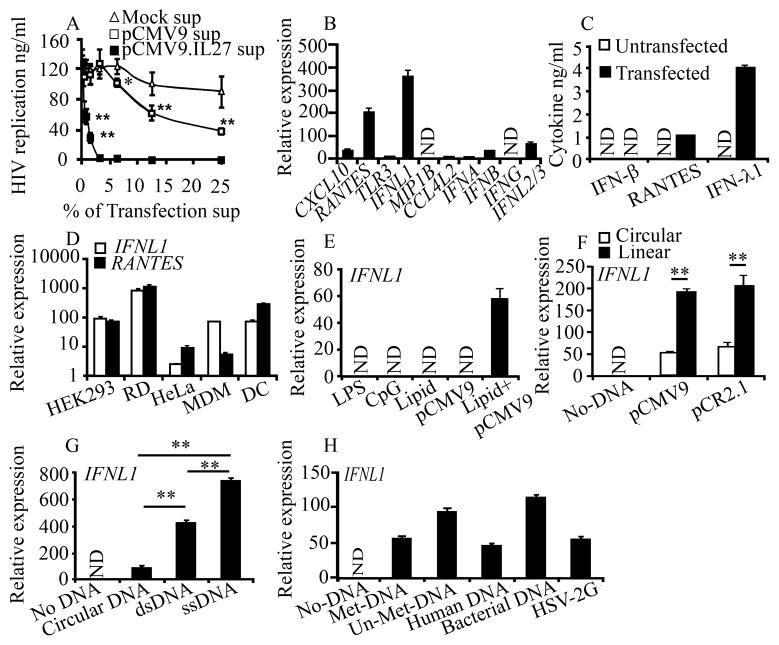
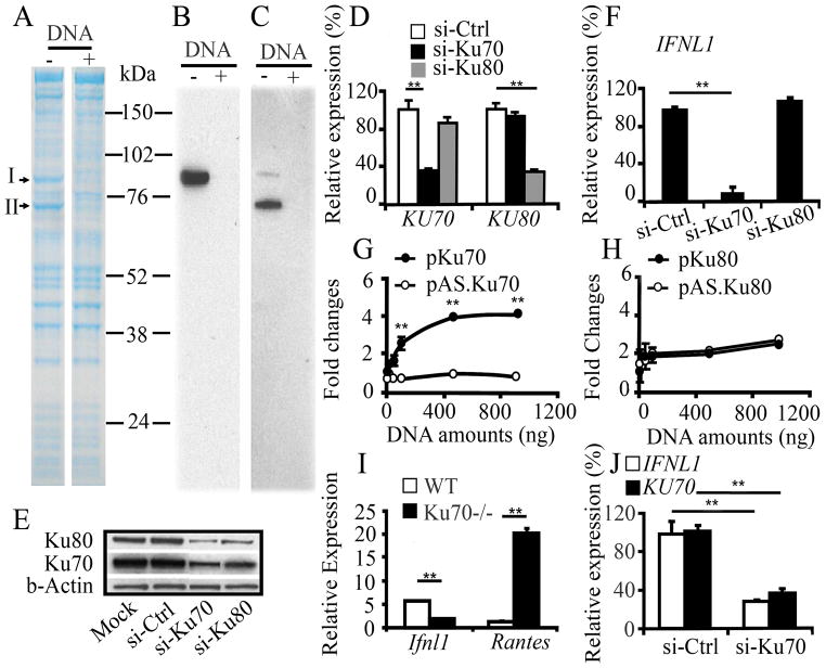
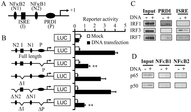
Cytosolic foreign DNA is detected by pattern recognition receptors and mainly induces type I IFN production. We found that transfection of different types of DNA into various untreated cells induces type III IFN (IFN-λ1) rather than type I IFN, indicating the presence of uncharacterized DNA sensor(s). A pull-down assay using cytosolic proteins identified that Ku70 and Ku80 are the DNA-binding proteins. The knockdown studies and the reporter assay revealed that Ku70 is a novel DNA sensor inducing the IFN-lambda1 activation. The functional analysis of IFNL1 promoter revealed that positive-regulatory domain I and IFN-stimulated response element sites are predominantly involved in the DNA-mediated IFNL1 activation. A pull-down assay using nuclear proteins demonstrated that the IFN-λ1 induction is associated with the activation of IFN regulatory factor-1 and -7. Thus, to our knowledge, we show for the first time that Ku70 mediates type III IFN induction by DNA.


Conflict of interest statement

Disclosures

The authors have no financial conflicts of interest.

Figures

Figure 1
Figure 1. Various types of DNA induce IFNL1 in different human cells
A, HIV-infected MDM were cultured with various concentrations (% v/v) of culture supernatants from pCMV9.IL27- or empty pCMV9-transfected HEK293 cells. HIV replication was determined using a p24 antigen capture assay. B, Gene expression in mock- or pCMV9- transfected HEK293 cells was confirmed by qRT-PCR. C, Cytokine concentrations in the transfection supernatants were determined via ELISA. DH, HEK293, HeLa, RD cells, MDM or DC cells were transfected with pCMV9 (D); HEK293 cells were treated with 100 ngml−1 LPS, 1 μM CpG ODN (InvivoGen), 3 μl of transfection lipid, 1 μg pCMV9 or a combination of the lipid and pCMV9 (E); HEK293 cells were treated with 1 μg supercoil (circular) or linearlized plasmids (pCMV9 and pCR2.1) (F); circular pCR2.1 (circular DNA), linearlized pCR2.1 (dsDNA), single-stranded pCR2.1 DNA (ssDNA) (G); linearized pCR2.1 (Met-DNA), PCR-amplified full-length pCR2.1 (Un-Met DNA), human DNA or bacterial DNA for 24 h, or infected with HSV-2G (MOI=5) for 18h (H), then gene expression was analyzed by qRT-PCR. Gene expression is presented as relative expression units compared with mocked transfection after normalization to GAPDH. Data is shown as the mean ± s.d. Data are mean ± s.d. (n=3), *p<0.05, ** p<0.01. N.D.: Not Detected.
Figure 2
Figure 2. Ku70 is a cytosolic DNA sensor positively regulating IFNL1 activation
A, Cytosol proteins from untreated HEK293 cells were incubated with DNA-conjugated beads in the absence or presence of DNA competitor (Supplemental Fig. 3). Proteins bound to the beads were separated on SDS–PAGE under reducing conditions, followed by Coomassie blue staining. B,C, Western blot analysis using anti-Ku70 (B) or anti-Ku80 (C) antibody demonstrated intended proteins. Due to a cross reactivity in the anti-Ku70 antibody, it detected Ku70 as well as Ku80. DE, HEK293 cells were transfected with si-Ctrl, si-Ku70 or si-Ku80, and the expression level of KU70 or KU80 mRNA (D) and protein (E) was analyzed by qRT-PCR and Western blot, respectively. The expression level of mRNA was compared with that in the cells transfected with si-Ctrl. Relative amounts of Ku70 and Ku80 protein levels were densitometrically analyzed using the NIH image, and normalized against β-actin. F, HEK293 cells were transfected with si-Ctrl, si-Ku70 or si-Ku80 followed by DNA transfection. Expression levels of IFNL1, KU70 and KU80 mRNA were determined by qRT-PCR. The level of mRNA was compared with that in the cells transfected with si-Ctrl. G–H, HEK293T cells were co-transfected with 100 ng full-length IFNL1–luciferase reporter plasmid and 10 ng Renilla luciferase plasmid with pKu70, pAS.Ku70, pKu80 or pAS.Ku80 for 24 h, then stimulated for 18 h by transfection with 500 ng of pCR2.1. I, Spleen cells from WT or KO Ku70−/− mice were transfected with linearized pCR2.1 using Nucleofactor Transfection kit, and then expression level of mRNA was analyzed by qRT-PCR. J, si-Ctrl or si-Ku70-transfected HEK293 cells were infected with HSV-2G, and then gene expression was analyzed. Data are shown as the mean ± s.d. (n = 3). **p<0.01.
Figure 3
Figure 3. PRDI and ISRE elements of the IFNL1 promoter are import for the DNA-mediated IFNL1 activation and IFNL1 activation is associated with the activation of IRF1 and 7
AB, Schematic representation of the IFNL1 promoter region and different mutant constructs on the IFNL1 promoter region. This diagram does not indicate the exact position of the elements. HEK293T cells were transfected with a series of variants of IFN-λ1–luciferase reporter and pTK-Ren for 24 h, and then stimulated with transfection of pCR2.1; the luciferase activities were normalized with Renilla activities and data are presented as fold inductions from promoter activity from basal promoter activation without pCR2.1 transfection. Data are shown as the mean ± s.d. (n = 3). **p<0.01. CD, Nuclear extracts from mock- or DNA-transfected HEK293 cells were allowed to bind to oligonucleotides (PRDI or ISRE or NFκB elements from the IFNL1 promoter) conjugated to beads. Proteins bound to the beads were separated on SDS-PAGE, followed by Western blot analysis with specific antibodies.

References

    1. Meylan E, Tschopp J, Karin M. Intracellular pattern recognition receptors in the host response. Nature. 2006;442:39–44. - PubMed
    1. Pichlmair A, Reis ESC. Innate recognition of viruses. Immunity. 2007;27:370–383. - PubMed
    1. Ranjan P, Bowzard JB, Schwerzmann JW, Jeisy-Scott V, Fujita T, Sambhara S. Cytoplasmic nucleic acid sensors in antiviral immunity. Trends Mol Med. 2009;15:359–68. - PubMed
    1. Takaoka A, Wang Z, Choi MK, Yanai H, Negishi H, Ban T, Lu Y, Miyagishi M, Kodama T, Honda K, Ohba Y, Taniguchi T. DAI (DLM-1/ZBP1) is a cytosolic DNA sensor and an activator of innate immune response. Nature. 2007;448:501–505. - PubMed
    1. Bird L. Innate immunity: Ready, AIM, fire! Nature reviews. 10:287. - PubMed

Publication types

MeSH terms