. 2011 Oct;45(4):858-66.
doi: 10.1165/rcmb.2010-0401OC. Epub 2011 Mar 11.

Novel murine model of chronic granulomatous lung inflammation elicited by carbon nanotubes

Affiliations

Novel murine model of chronic granulomatous lung inflammation elicited by carbon nanotubes

Isham Huizar et al. Am J Respir Cell Mol Biol. 2011 Oct.

Abstract

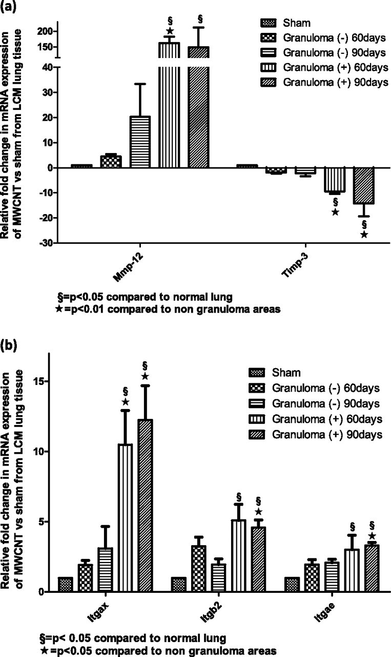
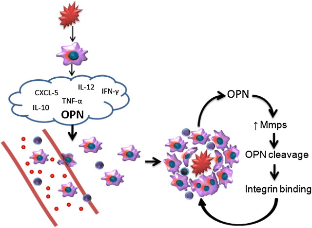
Lung granulomas are associated with numerous conditions, including inflammatory disorders, exposure to environmental pollutants, and infection. Osteopontin is a chemotactic cytokine produced by macrophages, and is implicated in extracellular matrix remodeling. Furthermore, osteopontin is up-regulated in granulomatous disease, and osteopontin null mice exhibit reduced granuloma formation. Animal models currently used to investigate chronic lung granulomatous inflammation bear a pathological resemblance, but lack the chronic nature of human granulomatous disease. Carbon nanoparticles are generated as byproducts of combustion. Interestingly, experimental exposures to carbon nanoparticles induce pulmonary granuloma-like lesions. However, the recruited cellular populations and extracellular matrix gene expression profiles within these lesions have not been explored. Because of the rapid resolution of granulomas in current animal models, the mechanisms responsible for persistence have been elusive. To overcome the limitations of previous models, we investigated whether a model using multiwall carbon nanoparticles would resemble chronic human lung granulomatous inflammation. We hypothesized that pulmonary exposure to multiwall carbon nanoparticles would induce granulomas, elicit a macrophage and T-cell response, and mimic other granulomatous disorders with an up-regulation of osteopontin. This model demonstrates: (1) granulomatous inflammation, with macrophage and T-cell infiltration; (2) resemblance to the chronicity of human granulomas, with persistence up to 90 days; and (3) a marked elevation of osteopontin, metalloproteinases, and cell adhesion molecules in granulomatous foci isolated by laser-capture microdissection and in alveolar macrophages from bronchoalveolar lavage. The establishment of such a model provides an important platform for mechanistic studies on the persistence of granuloma.


Figures

Figure 1.
Figure 1.
(a) Diameter distribution of multiwall carbon nanotubes (MWCNTs) obtained from scanning electron microscopy images in c and d. (b) Raman spectrum of MWNTs reveals presence of a D peak (∼1,350 cm−1), G peak (∼1,580 cm−1), and D′ peak (1,620 cm−1). (c and d) Typical scanning electron microscopy images of MWCNTs.
Figure 2.
Figure 2.
Persistence of granulomatous inflammation for up to 90 days after exposure to MWCNTs. Hematoxylin and eosin staining was applied to (a) murine lung control tissue and to murine lung tissue (b) 10 days, (c) 60 days, and (d) 90 days after exposure to MWCNTs. The granulomatous reaction surrounding MWCNT aggregates begins 10 days after exposure, and persists for 60 and 90 days after exposure to MWCNTs, without apparent resolution.
Figure 3.
Figure 3.
The expression of osteopontin mRNA is up-regulated in granulomatous foci. (a) Osteopontin gene expression of lung tissue obtained through laser-capture microdissection (LCM) in sham untreated mice, surrounding normal lung tissue [Granuloma (−)] and granulomatous foci [Granuloma (+)]. The recruitment of CD3+ T cells and macrophages, with fusion and multinucleated giant cells, is evident. Immunohistochemistry for macrophages (monocyte + macrophage [MOMA]; green) demonstrates macrophages surrounding MWCNT aggregates, (b) 60 days and (c) 90 days after exposure to MWCNTs. (d and e) Immunohistochemistry for CD3+ T cells (red) and osteopontin (green) found within granulomatous lesions of murine lungs, (d) 60 days and (e) 90 days after exposure to MWCNTs. (f and g) Immunohistochemistry for CD3+ T cells (red) and CD4+ (green) in murine lungs, (f) 60 days and (g) 90 days after exposure to MWCNTs. Arrows indicate locations of MWCNT aggregates.
Figure 4.
Figure 4.
Osteopontin is increased in bronchoalveolar lavage (BAL) from mice exposed to MWCNTs at 60 and 90 days. (a) mRNA gene expression for matrix metalloproteinases (MMPs) in BAL, 60 and 90 days after exposure to MWCNTs. (b) Osteopontin protein concentrations in BAL, 60 and 90 days after exposure to MWCNTs. (c) Cytospin of BAL 90 days after exposure to MWCNTs reveals a macrophage-containing MWCNT within the cytoplasm. (d–f) Immunohistochemistry stain of BAL for macrophage marker MOMA (green), osteopontin (red), and overlap (orange) in (d) control mice, and (e) 60 days and (f) 90 days after exposure to MWCNTs.
Figure 5.
Figure 5.
(a) Granulomatous foci are associated with an up-regulation in matrix proteases and a down-regulation of tissue inhibitor of metalloproteinases (TIMP)–3. The mRNA gene expression of MMPs and TIMP-3 was obtained from a PCR gene array of lung tissue via LCM from sham untreated mice, unaffected lung tissue [Granuloma (−)], and granulomatous foci [Granuloma (+)]. (b) Integrins (Itgs) are elevated within granulomatous foci at 60 and 90 days after exposure to MWCNTs. The mRNA gene expression of integrins was obtained from a PCR gene array of lung tissue obtained via LCM from sham untreated mice, unaffected lung [Granuloma (−)], and granulomatous foci [Granuloma (+)]. Itgs are elevated within granulomatous foci at 60 and 90 days after exposure to MWCNTs.
Figure 6.
Figure 6.
Proposed mechanisms for the formation of granulomas. The initial insult causes a release of cytokines, with subsequent recruitment, attachment, and transformation of macrophages and T cells. Osteopontin (OPN) within granulomas is proteolytically cleaved by MMPs binding to integrins and enabling macrophage fusion. These events result in the continued expression of osteopontin and MMPs, with subsequent transformation of macrophages into epithelioid and multinucleated giant cells, and the retention of T cells within granulomatous foci. CXCL, chemokine (C-X-C motif) ligand 1.

References

    1. Helming L, Gordon S. The molecular basis of macrophage fusion. Immunobiology 2007;212:785–793. - PubMed
    1. Castranova V. Signaling pathways controlling the production of inflammatory mediators in response to crystalline silica exposure: role of reactive oxygen/nitrogen species. Free Radic Biol Med 2004;37:916–925. - PubMed
    1. Baughman RP, Lower EE. du Bois RM. Sarcoidosis. Lancet 2003;361:1111–1118. - PubMed
    1. Pagnoux C. Churg-Strauss syndrome: evolving concepts. Discov Med 2010;9:243–252. - PubMed
    1. Wieczorek S, Holle JU, Epplen JT. Recent progress in the genetics of Wegener's granulomatosis and Churg-Strauss syndrome. Curr Opin Rheumatol 2010;22:8–14. - PubMed

Publication types

MeSH terms