. 2011 Jul;39(12):5045-56.
doi: 10.1093/nar/gkr099. Epub 2011 Mar 11.

PARG is recruited to DNA damage sites through poly(ADP-ribose)- and PCNA-dependent mechanisms

Affiliations

PARG is recruited to DNA damage sites through poly(ADP-ribose)- and PCNA-dependent mechanisms

Oliver Mortusewicz et al. Nucleic Acids Res. 2011 Jul.

Abstract

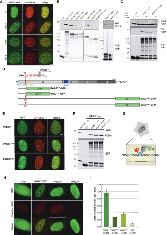
Post-translational poly(ADP-ribosyl)ation has diverse essential functions in the cellular response to DNA damage as it contributes to avid DNA damage detection and assembly of the cellular repair machinery but extensive modification eventually also induces cell death. While there are 17 human poly(ADP-ribose) polymerase (PARP) genes, there is only one poly(ADP-ribose) glycohydrolase (PARG) gene encoding several PARG isoforms located in different subcellular compartments. To investigate the recruitment of PARG isoforms to DNA repair sites we locally introduced DNA damage by laser microirradiation. All PARG isoforms were recruited to DNA damage sites except for a mitochondrial localized PARG fragment. Using PARP knock out cells and PARP inhibitors, we showed that PARG recruitment was only partially dependent on PARP-1 and PAR synthesis, indicating a second, PAR-independent recruitment mechanism. We found that PARG interacts with PCNA, mapped a PCNA binding site and showed that binding to PCNA contributes to PARG recruitment to DNA damage sites. This dual recruitment mode of the only nuclear PARG via the versatile loading platform PCNA and by a PAR dependent mechanism likely contributes to the dynamic regulation of this posttranslational modification and ensures the tight control of the switch between efficient DNA repair and cell death.


Figures

Figure 1.
Figure 1.
Recruitment of PARG isoforms to laser-induced DNA damage sites. (A) Schematic representation of PARG and fusion proteins used. (B) Live cell imaging of a microirradiated HeLa cell expressing PARG111-GFP. Comparision of PARG111-GFP (C), GFP-PARP-1 (D) and GFP-XRCC1 (E) recruitment to DNA damage sites in HeLa cells and quantitative evaluation of recruitment kinetics showing mean curves. Recruitment of the PARG isoforms PARG111-GFP (F), PARG102-GFP (G) and PARG99-GFP (H) to laser-induced DNA damage sites in mouse embryonic fibroblasts (MEFs). (I) The mitochondrial isoform PARG461-976-GFP shows no accumulation at microirradiated sites. Where no recruitment can be observed, sites of irradiation are indicated by arrows. Error bars represent the standard error of the mean. Scale bar: 5 µm.
Figure 2.
Figure 2.
PAR-dependent recruitment of PARG to DNA damage sites. (A) Schematic representation of PARG and fusion proteins used. (B) Live cell imaging of PARG111-GFP recruitment in wt MEFs (upper row), Parp-1−/− MEFs (middle row) and in wt MEFs treated with the PARP inhibitor NU1025 (lower row). (C) Comparison of PARG111-GFP recruitment kinetics in wt MEFs (green curve), Parp-1−/− MEFs (red curve) and wt MEFs treated with NU1025 (blue curve). (D) Quantitative evaluation of FRAP data showing mean curves. To analyse the mobility of PARG111-GFP at DNA damage sites in wt MEFs (green curve), Parp-1−/− MEFs (red curve) and wt MEFs treated with NU1025 (blue curve) the microirradiated region was bleached 5 min after microirradiation and the fluorescence recovery was measured. (E) Accumulation of the N-terminal domain of PARG (PARG1–469-GFP) at laser-induced DNA damage sites. (F) Quantitative evaluation of PARG1–469-GFP recruitment kinetics in wt MEFs (green curve), Parp-1−/− MEFs (red curve) and wt MEFs treated with NU1025 (blue curve) showing mean curves. (G) The C-terminal domain of PARG (PARG478–976-GFP) shows no accumulation at DNA damage sites. Where no recruitment can be observed, sites of irradiation are indicated by arrows. Error bars represent the standard error of the mean. Scale bar: 5 µm.
Figure 3.
Figure 3.
Inactive PARG is recruited with higher efficiency to laser-induced DNA damage sites. (A) Schematic representation of PARG and PARGE755,756A-GFP. Mutated amino acid positions are indicated in red. (B) Zymogram showing the PAR-degrading capacity of the indicated GFP-tagged PARG proteins. (C) Live cell imaging of PARGE755,756A-GFP recruitment in wt MEFs (upper row), Parp-1−/− MEFs (middle row) and in wt MEFs treated with the PARP inhibitor NU1025 (lower row). (D) Recruitment kinetics of PARGE755,756A-GFP in wt MEFs (green curve), Parp-1−/− MEFs (red curve) and wt MEFs treated (blue curve) or not (black curves) with NU1025. (E) Mobility of PARGE755,756A-GFP at DNA damage sites in wt MEFs (green curve), Parp-1−/− MEFs (red curve) and wt MEFs treated with NU1025 (blue curve). (F) Immunodetection of PAR in PARGKD cells expressing shRNA-insensitive PARGE755,756A-GFP or PARG111-GFP, at the time indicated after 10 min of treatment with 0.5 mM H2O2. Error bars represent the standard error of the mean. Scale bar: 5 µm.
Figure 4.
Figure 4.
PARG interacts with PCNA and is present at replication foci. (A) Colocalization of PARG with PCNA throughout S-phase. Spinning disk microscopy images of wt MEFs coexpressing PARG111-GFP and RFP-PCNA in different S-phase stages. PARG111-GFP colocalizes with RFP-PCNA at sites of replication throughout S-phase. (B) Interaction of PARG with PCNA analysed by GFP-pull down assays. The indicated GFP-tagged proteins were overexpressed in HEK293T cells, recovered by GFP-pulldown and copurified PCNA was assessed by western blot with anti PCNA antibodies (upper panels). The presence of GFP-tagged protein is revealed with an anti GFP antibody (lower panels). (C) Effect of H2O2 treatment on the interaction between PARG and PCNA. PARG111-GFP and PARGE755,756A-GFP overexpressing HEK293T cells were treated with 1 mM H2O2 for 10 min before performing a GFP-pull down assay as described in (B). The asterisk indicates mono-ubiquitinated PCNA, visible in the input of H2O2-treated cells (1% input was loaded on the gel). (D) Schematic representation of PARG including the identified PIP box sequence and fusion proteins used. Mutated amino acid positions are indicated in red. (E) Spinning disk microscopy images of wt MEFs expressing PARG111-GFP, PARGQ76A-GFP or PARGQ76E-GFP and stained with a mouse-monoclonal antibody against PCNA to mark sites of replication. (F) Effect of the mutation of the PARG PIP domain on its interaction with PCNA. GFP-pull down assays with the indicated PARG constructs were performed as in (B). (G) Schematic outline of the F2H assay (see text for details). (H) U2OS.2-6-3 cells with a stably integrated lac operator array coexpressing PCNA-LacI-RFP (bait protein) and various GFP-tagged PARG fusions (prey proteins). Colocalization of the red PCNA-LacI-RFP signal with a green prey signal at the lac operator array indicates interaction. (I) Quantification of the relative enrichment of PARG111-GFP, PARGQ76A-GFP, PARGQ76E-GFP and GFP at the lac operator array. For quantification, the fluorescence signal at the lac operator array was measured and divided by the overall fluorescence measured in the nucleus. Error bars represent the standard error of the mean. Scale bar: 5 µm.
Figure 5.
Figure 5.
PARG interaction with PCNA contributes to its accumulation at laser-induced DNA damage sites. (A) Accumulation and recruitment kinetics of PARG1-87-GFP to laser-induced DNA damage sites. The recruitment kinetics of PARG1-87-GFP in wt MEFs (green curve), Parp-1−/− MEFs (red curve) and wt MEFs treated with NU1025 (blue curve) as well as the recruitment kinetics for PARG111-GFP in wt MEFs (light green) are shown for comparison. (B) PARG1-75-GFP lacking the newly identified PIP box does not accumulate at DNA damage sites. (C) Mutation of the newly identified PIP box of PARG (PARGQ76A-GFP) decreases PARG accumulation to DNA damage sites. Recruitment kinetics of PARGQ76A-GFP in wt MEFs (green curve), Parp-1−/− MEFs (red curve) and wt MEFs treated with NU1025 (blue curve) as well as the recruitment kinetics for PARG111-GFP in wt MEFs (light green) are shown for comparison. (D) Comparison of PARGQ76A-GFP (dark blue curve) and PARG111-GFP (light blue curve) recruitment kinetics in wt MEFs treated with NU1025. Error bars represent the standard error of the mean. Scale bar: 5 µm. (E) Live cell imaging of cherry-PCNA recruitment in wt MEFs (upper row), Parp-1−/− MEFs (middle row) and in wt MEFs treated with the PARP inhibitor NU1025 (lower row). (F) Comparison of cherry-PCNA recruitment kinetics in wt MEFs (red curve), Parp-1−/− MEFs (blue curve) and wt MEFs treated with NU1025 (green curve). Error bars represent the standard error of the mean. Where no recruitment can be observed, sites of irradiation are indicated by arrows. Scale bar: 5 µm. (G) PCNA does not bind PAR on polymer blot assay. Recombinant purified BSA, histone H1 and PCNA were spotted on nitrocellulose membrane as indicated before incubation with 32P-labelled PAR (right panel). Same amounts of proteins were analysed by SDS–PAGE gel electrophoresis and Coomassie staining to control the quality and quantities of the proteins used in the assay.

Similar articles

Cited by

References

    1. Hakmé A, Huber A, Dollé P, Schreiber V. The macroPARP genes Parp-9 and Parp-14 are developmentally and differentially regulated in mouse tissues. Dev. Dyn. 2008;237:209–215. - PMC - PubMed
    1. Poirier GG, de Murcia G, Jongstra-Bilen J, Niedergang C, Mandel P. Poly(ADP-ribosyl)ation of polynucleosomes causes relaxation of chromatin structure. Proc. Natl Acad. Sci. USA. 1982;79:3423–3427. - PMC - PubMed
    1. El-Khamisy SF, Masutani M, Suzuki H, Caldecott KW. A requirement for PARP-1 for the assembly or stability of XRCC1 nuclear foci at sites of oxidative DNA damage. Nucleic Acids Res. 2003;31:5526–5533. - PMC - PubMed
    1. Mortusewicz O, Ame JC, Schreiber V, Leonhardt H. Feedback-regulated poly(ADP-ribosyl)ation by PARP-1 is required for rapid response to DNA damage in living cells. Nucleic Acids Res. 2007;35:7665–7675. - PMC - PubMed
    1. Okano S, Lan L, Caldecott KW, Mori T, Yasui A. Spatial and temporal cellular responses to single-strand breaks in human cells. Mol. Cell. Biol. 2003;23:3974–3981. - PMC - PubMed

Publication types

Substances