. 2011 Apr 14;472(7342):230-3.
doi: 10.1038/nature09932. Epub 2011 Mar 13.

Structure of mammalian AMPK and its regulation by ADP

Affiliations

Structure of mammalian AMPK and its regulation by ADP

Bing Xiao et al. Nature. .

Abstract

The heterotrimeric AMP-activated protein kinase (AMPK) has a key role in regulating cellular energy metabolism; in response to a fall in intracellular ATP levels it activates energy-producing pathways and inhibits energy-consuming processes. AMPK has been implicated in a number of diseases related to energy metabolism including type 2 diabetes, obesity and, most recently, cancer. AMPK is converted from an inactive form to a catalytically competent form by phosphorylation of the activation loop within the kinase domain: AMP binding to the γ-regulatory domain promotes phosphorylation by the upstream kinase, protects the enzyme against dephosphorylation, as well as causing allosteric activation. Here we show that ADP binding to just one of the two exchangeable AXP (AMP/ADP/ATP) binding sites on the regulatory domain protects the enzyme from dephosphorylation, although it does not lead to allosteric activation. Our studies show that active mammalian AMPK displays significantly tighter binding to ADP than to Mg-ATP, explaining how the enzyme is regulated under physiological conditions where the concentration of Mg-ATP is higher than that of ADP and much higher than that of AMP. We have determined the crystal structure of an active AMPK complex. The structure shows how the activation loop of the kinase domain is stabilized by the regulatory domain and how the kinase linker region interacts with the regulatory nucleotide-binding site that mediates protection against dephosphorylation. From our biochemical and structural data we develop a model for how the energy status of a cell regulates AMPK activity.


Figures

Figure 1
Figure 1. Role of ADP in regulation of AMPK activity
(a) AMP, but not ADP, allosterically activates AMPK. (b) AMP and ADP protection of AMPK from dephosphorylation. (c) ATP does not protect against dephosphorylation. (d) Mg.ATP competes with the protective effect of ADP on dephosphorylation. Results are displayed as the mean ± S.E.M determined from at least 3 independent experiments. Where appropriate a representative blot (n=3) showing Thr-172 phosphorylation and total α subunit levels is shown.
Figure 2
Figure 2. Measurement of equilibrium dissociation constants for the binding of AXPs to phosphorylated AMPK
(a) Displacement of Coumarin-ATP from the AMPK:(Coumarin-ATP)2 complex by AXPs monitored using fluorescence at 470 nm. Solid lines are the computed best fits with Kd,I and Kd,II for C-ATP binding to AMPK fixed at 1.1 and 4.2 μM. Inset: Titration of Coumarin-ATP with AMPK. (b) Displacement of NADH from the AMPK:NADH complex by AXPs monitored using fluorescence at 435 nm. The solid line is the computed best fit with the Kd for NADH fixed at 65 μM. Inset: Fluorescence titration of NADH with AMPK.
Figure 3
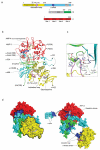
Figure 3. Crystal structure of active mammalian AMPK
(a) Schematic representation of the components of the heterotrimer; the parts of the complex missing from the crystallized protein are shown in grey. The domains, including the activation loop (pink) and α-hook (dark blue), are coloured the same in all panels. (b) Ribbon representation of the crystallized complex with two the bound AMPs, staurosporine and phospho Thr-172 shown in stick representation. The α-hook and activation loop of the kinase domain are shown in heavier lines and coloured dark blue and pink respectively. (c) The interface between the activation loop and the regulatory fragment are shown in more detail in a similar orientation as (b), potential electrostatic interactions are indicated by dashed lines. (d) The complex is shown in two space-filling representations. The left panel represents the same view as (b) with the α-hook and kinase domain outlined in black. In the right hand panel these two components have been rotated away from the regulatory domain to show the interaction surfaces in an ‘open-book’ representation, where the contacting residues have been coloured in dark blue. With the α-hook removed, AMP-3 becomes visible. The black arrows indicate the rotations that would reassemble the complex.
Figure 4
Figure 4. Mutational analysis of AMPK regulation
(a) Desphophorylation rate of the wild-type (WT) or β His-233 to alanine kinase domain interface mutant (H233A, corresponding to H235A in β2). (b) Protection of dephosphorylation of WT or H233A mutant by AMP (30 μM) or ADP (30 μM) after incubation for 5 mins. (c) Allosteric activation of WT or α-hook mutant (harbouring mutation of residues R375Q, T377A, D379A and E380A within α1) by AMP (100 μM). (d) AMP (30 μM) and ADP (30 μM) protection of WT or α-hook mutant from dephosphorylation. Results are the mean ± S.E.M from at least 3 independent experiments

Comment in

  • Signal transduction: How cells sense energy.
    Hardie DG. Hardie DG. Nature. 2011 Apr 14;472(7342):176-7. doi: 10.1038/472176a. Nature. 2011. PMID: 21490664 No abstract available.
  • Conserved regulatory elements in AMPK.
    Chen L, Xin FJ, Wang J, Hu J, Zhang YY, Wan S, Cao LS, Lu C, Li P, Yan SF, Neumann D, Schlattner U, Xia B, Wang ZX, Wu JW. Chen L, et al. Nature. 2013 Jun 13;498(7453):E8-10. doi: 10.1038/nature12189. Nature. 2013. PMID: 23765502 No abstract available.

References

    1. Carling D. The AMP-activated protein kinase cascade--a unifying system for energy control. Trends Biochem Sci. 2004;29:18–24. - PubMed
    1. Hardie DG. AMP-activated/SNF1 protein kinases: conserved guardians of cellular energy. Nat Rev Mol Cell Biol. 2007;8:774–785. - PubMed
    1. Kahn BB, Alquier T, Carling D, Hardie DG. AMP-activated protein kinase: ancient energy gauge provides clues to modern understanding of metabolism. Cell Metab. 2005;1:15–25. - PubMed
    1. Shackelford DB, Shaw RJ. The LKB1-AMPK pathway: metabolism and growth control in tumour suppression. Nat Rev Cancer. 2009;9:563–575. - PMC - PubMed
    1. Cool B, et al. Identification and characterization of a small molecule AMPK activator that treats key components of type 2 diabetes and the metabolic syndrome. Cell Metab. 2006;3:403–416. - PubMed

Publication types

MeSH terms