. 2011 Jun;63(6):1613-25.
doi: 10.1002/art.30311.

Transcriptome-wide gene regulation by gentle treadmill walking during the progression of monoiodoacetate-induced arthritis

Affiliations

Transcriptome-wide gene regulation by gentle treadmill walking during the progression of monoiodoacetate-induced arthritis

Jin Nam et al. Arthritis Rheum. 2011 Jun.

Abstract

Objective: Physiotherapies are the most widely recommended conservative treatment for arthritic diseases. The present study was undertaken to examine the molecular mechanisms underlying the effects of gentle treadmill walking (GTW) on various stages of monoiodoacetate-induced arthritis (MIA) to elucidate the basis for the success or failure of such therapies in joint damage.

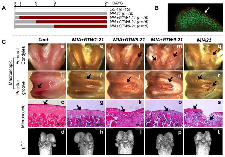
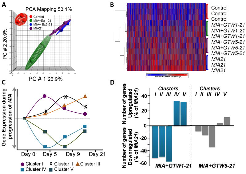
Methods: Knees were obtained from untreated control rats, rats with MIA that did not undergo GTW, rats with MIA in which GTW regimens were started 1 day post-MIA induction, and rats with MIA in which GTW regimens were started after cartilage damage had progressed to grade 1 or grade 2. The cartilage was examined macroscopically, microscopically, and by microfocal computed tomography imaging. Transcriptome-wide gene expression analysis was performed, and microarray data were assessed by Ingenuity Pathways Analysis to identify molecular functional networks regulated by GTW.

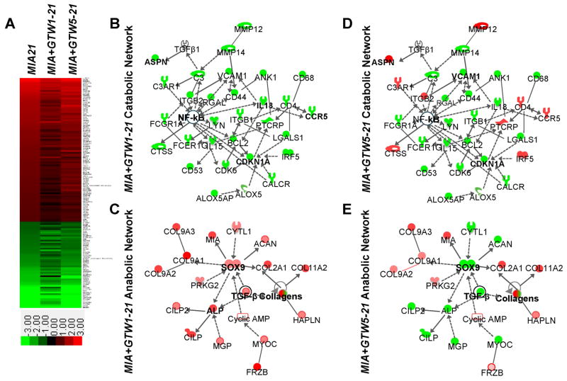
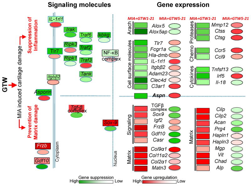
Results: GTW intervention started on day 1 post-MIA induction significantly prevented the progression of MIA, but its efficacy was reduced when implemented on knees exhibiting close to grade 1 cartilage damage. GTW accelerated cartilage damage in knees with close to grade 2 damage. Transcriptome-wide gene expression analysis revealed that GTW intervention started 1 day post-MIA inception significantly suppressed inflammation-associated genes and up-regulated matrix-associated gene networks. However, delayed GTW intervention after grade 1 damage had occurred was less effective in suppressing proinflammatory genes or up-regulating matrix synthesis.

Conclusion: The present findings suggest that GTW suppresses proinflammatory gene networks and up-regulates matrix synthesis to prevent progression of cartilage damage in MIA-affected knees. However, the extent of cartilage damage at the initiation of GTW may be an important determinant of the success or failure of such therapies.


Conflict of interest statement

All authors did not have any other financial interests that may create a potential conflict of interest or the appearance of a conflict of interest with regard to the present work.

Figures

Figure 1
Figure 1
Effects of GTW on the progression of MIA. (A) Experimental scheme. (B) Cartilage section showing dead (red) and live (green) cells on day 1 post-MIA inception. (C) Macroscopic, microscopic and bone imaging by μCT of: healthy sham control femur showing smooth surface, normal histology and no bone lesions on the femoral condyles and patellar grove (see Supplemental Data for 360° μCT projections) (a – d); MIA+GTW1-21 cartilage showing no surface abrasions on the condyles, some cartilage lesions on the patellar groove and ridges, near normal histology, and no bone involvement by μCT images (e – h); MIA+GTW5-21 cartilage showing some abrasions on condyles, cartilage damage on patellar groove and ridges, H&E section showing focal matrix condensation, cell clustering and disorganization, fibrocartilage formation, and some bone lesions by μCT images (i – l); MIA+GTW9-21 cartilage demonstrating extensive cartilage lesions on condyles and patellar groove, severe cartilage loss, denuded bone, and excessive bone lesions on femoral condyles and patellar grove in μCT images (m – p); MIA21 cartilage exhibiting cartilage matrix loss, delamination of superior surface, excavation and matrix loss in superficial and mid zone (q – t). Each femur is representative of each group showing similar characteristics (n=10).
Figure 2
Figure 2
Transcriptome-wide microarray analysis of femoral cartilage from Cont healthy joints or from MIA+GTW1-21, MIA+GTW5-21 or MIA21 joints. (A) PCA analysis showing reproducible gene expression in the articular cartilage from the right knee joint of 3 separate rats from Cont, MIA+Ex1-21, MIA+GTW5-21 or MIA21. (B) Overall gene expression profiles of the articular cartilage of 3 separate rats from Cont, MIA+GTW1-21, MIA+GTW5-21 or MIA21 groups. Intensity plot with dendrogram represents the transcripts that were significantly (p < 0.05) and differentially upregulated (red) or downregulated (blue) by more than two-fold. The analysis shows the most differential changes in gene expression in MIA+GTW1-21, followed by MIA+GTW5-21 as compared to gene expression in MIA21 cartilage. (C) Temporal regulation of MIA associated Gene Clusters associated with: acute and innate immune responses (Cluster I); chronic inflammatory responses and genetic disorders (Cluster II); musculoskeletal disorders and inflammatory diseases (Cluster III); and genetic disorders and skeletal and muscular disorders (Cluster IV and V). (D) The percentage of genes that were significantly (p<0.05) up or downregulated by exercise (MIA+GTW1-21 and MIA+GTW5-21) in gene Clusters I, II, III, IV and V in comparison to genes regulated in MIA21.
Figure 3
Figure 3
Catabolic and anabolic networks regulated by GTW in MIA. (A) Intensity plot of 142 genes that are a known set of arthritis-associated genes in Ingenuity Knowledge Database, and more than 2-fold up (red) or down (green) regulated in MIA21 (see Supplemental Data for details). (B–E) IPA generated networks of: (B) catabolic genes that are downregulated by GTW in MIA+GTW1-21 showing suppression of genes involved in acute and chronic inflammatory/immune responses via the suppression of NF-κB activity; (C) anabolic genes that are upregulated by exercise in MIA+GTW1-21 showing induction of Sox9 and TGF-β that in turn may upregulate the expression of matrix associated genes; (D) catabolic gene regulation by GTW in MIA+GTW5-21 showing partial suppression of genes involved in acute and chronic immune responses; (E) anabolic gene regulation by GTW in MIA+GTW5-21 showing suppression of Sox9 and TGF-β and number of matrix proteins that may limit its ability to repair cartilage. Red, green, and white colors represent upregulation, downregulation and no regulation, respectively. The shading of colors represents dark, greater changes to light lesser changes. Symbols are: formula image cytokine/growth factor, formula image phosphatase, formula image Transcription regulator, formula image translation regulator, formula image transmembrane receptor, formula image complex group, formula image enzyme, formula image G protein coupled receptor, formula image kinase, formula image peptidase, and formula image other.
Figure 4
Figure 4
(A) Genes selected from each Cluster to demonstrate their up- or downregulation by quantitative rt-PCR analysis in MIA+GTW1-21 or MIA+GTW5-21 cartilage, as compared to MIA21 cartilage. Fcgr1a (IgG Fc receptor gamma 1a), Aspn (Asporin), Mmp12 (Matrix metalloproteinase 12), Alox5 (Arachidonate-5-lipoxygenase), Vcam1 (vascular endothelial cell adhesion molecule 1), Cilp (cartilage intermediate layer protein), Sox9 (Sry-related HMG box -9), Col9α1 (Collagen type 9α1), Frzb (Frizzled B), Col2α1 (Collagen type 2α1). Data represents rt-PCR analysis performed on RNA from five separate rats in each group. The * and ** indicate significance values of p > 0.05 and p > 0.01, respectively. (B) Regulation of the genes required for the activation of NF-κB by GTW. Suppression of Traf2 (TNF receptor associated factor), Traf3, Traf6, Tank (TRAF family member-associated NF-kappa-B activator), Ripk1 (Receptor (TNFRSF) interacting Ser-Thr kinase), Ripk3, Ikbkg (I-κb kinase γ/IKK γ) expression in MIA+GTW1-21and MIA+GTW5-21, as compared to MIA21 in the microarray data. Upregulation of the gene expression in MIA21 was compared to Cont. Each point represents mean of microarray data derived from 3 independent cartilage specimens (p<0.05).
Figure 5
Figure 5
Schematic showing the molecular basis of the protective effects of GTW on MIA-induced cartilage damage. GTW preserves cartilage integrity by preventing MIA-induced inflammation and matrix loss in MIA+GTW1-21 (rectangles) to a greater extent than in MIA+GTW5-21 (ovals), where GTW was applied following Grade 1 cartilage damage. These differential effects stem from the ability of GTW to suppress (green) MIA-induced NF-κB signaling molecules in MIA+GTW1-21, as compared to minimal suppression or upregulation (red) of these molecules in MIA+GTW5-21. This in turn may suppress several proinflammatory genes [arachidonate metabolites (Arach), receptors, proteases, chemokines (Chemo) and cytokines] in MIA+GTW1-21, but to a lesser degree in MIA+GTW5-21. Importantly, Aspn, an inhibitor of TGF-β that is upregulated in MIA21, is significantly suppressed by GTW in MIA+GTW1-21. By suppressing Asporin expression, GTW may upregulate TGF-β complex and SOX-9 in MIA+GTW1-21, required for synthesis of matrix proteins. Simultaneous upregulation of Frizzled-related protein (Frzb) likely inhibit chondrocyte hypertrophy and mineralization (50). Contrarily, the lack of Asporin suppression and thus failure of the expression of the molecules in TGF-β complex and Sox-9 in MIA+GTW5-21 may prevent MIA-induced matrix loss to a lesser extent, as evidenced by the suppression of several molecules required for matrix assembly.

References

    1. Hootman JM. Osteoarthritis in elderly persons: risks of exercise and exercise as therapy. Clin J Sport Med. 2010;20:223. - PubMed
    1. Hunter DJ, Lo GH. The management of osteoarthritis: an overview and call to appropriate conservative treatment. Med Clin North Am. 2009;93:127–43. xi. - PubMed
    1. Zhang W, Nuki G, Moskowitz RW, Abramson S, Altman RD, Arden NK, et al. OARSI recommendations for the management of hip and knee osteoarthritis: part III: Changes in evidence following systematic cumulative update of research published through January 2009. Osteoarthritis Cartilage. 2010;18:476–99. - PubMed
    1. Dunlop DD, Semanik P, Song J, Sharma L, Nevitt M, Jackson R, et al. Moving to maintain function in knee osteoarthritis: evidence from the osteoarthritis initiative. Arch Phys Med Rehabil. 2010;91:714–21. - PMC - PubMed
    1. Penninx BW, Messier SP, Rejeski WJ, Williamson JD, DiBari M, Cavazzini C, et al. Physical exercise and the prevention of disability in activities of daily living in older persons with osteoarthritis. Arch Intern Med. 2001;161:2309–16. - PubMed

Publication types

Substances