. 2011 Apr;69(4):681-90.
doi: 10.1002/ana.22278. Epub 2011 Mar 11.

Alternative splicing dysregulation secondary to skeletal muscle regeneration

Affiliations

Alternative splicing dysregulation secondary to skeletal muscle regeneration

James P Orengo et al. Ann Neurol. 2011 Apr.

Abstract

Objective: Dysregulation of alternative splicing has become a molecular hallmark of myotonic dystrophy type 1 (DM1), in which neonatal splice variants are expressed in adult skeletal muscle. Splicing dysregulation is induced by RNA containing expanded CUG repeats expressed from the expanded mutant allele by sequestration of muscleblindlike 1 (MBNL1) protein within nuclear RNA foci and increased CUGBP, ELAV-like family member 1 (CELF1) protein levels. Dysregulated splicing has also been identified in other neuromuscular disorders, suggesting either that diseases with different molecular causes share a common pathogenic mechanism or that dysregulated splicing can also be a common secondary consequence of muscle degeneration and regeneration.

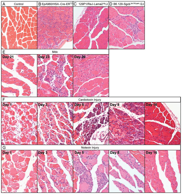
Methods: In this study, we examined regulation of alternative splicing in 4 different mouse models of muscular dystrophy, including DM1, limb-girdle muscular dystrophy, congenital merosin-deficient muscular dystrophy, and Duchenne muscular dystrophy, and 2 myotoxin (cardiotoxin and notexin) muscle injury models.

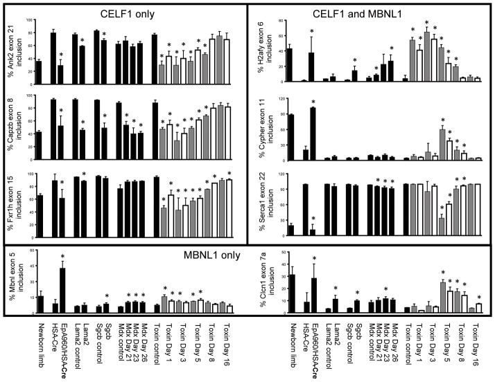
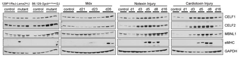
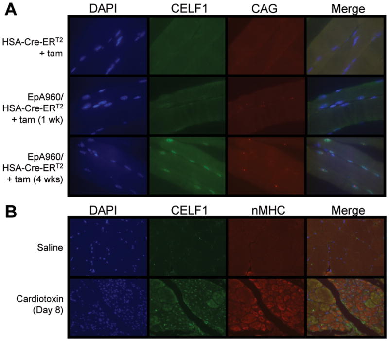
Results: We show that DM1-like alternative splicing dysregulation and altered expression of MBNL1 and CELF1 occur in non-DM1 mouse models of muscular dystrophy and muscle injury, most likely due to recapitulation of neonatal splicing patterns in regenerating fibers. In contrast, CELF1 was elevated in nuclei of mature myofibers of the DM1 model, consistent with a primary effect of pathogenic RNA expression.

Interpretation: Splicing dysregulation in DM1 is a primary effect of RNA containing expanded CUG repeats. However, we conclude that splicing changes can also be observed secondary to muscle regeneration, and this possibility must be taken into account when evaluating cause-effect relationships between dysregulated splicing and disease processes.


Figures

Figure 1
Figure 1. Histological analysis of six models of muscular dystrophy and muscle injury
Hematoxylin and eosin stained cross-sections from paraffin embedded gastrocnemius muscle at 20X magnification. (A) Wild-type control, (B) EpA960/HSA-Cre-ERT2 (+ tam), (C) 129P1/ReJ-Lama2dy/J, (D) B6.129-Sgcbtm1Kcam/2J, (E) mdx at postnatal days 21, 23 and 26, (F) cardiotoxin and (G) notexin injured muscle at 1, 3, 5, 8 and 16 days post injection.
Figure 2
Figure 2. Splicing misregulation in several muscular dystrophy and muscle injury models
RT-PCR was performed on RNA extracted from gastrocnemius muscle using primers that anneal to constitutive exons flanking the alternative exon (Supp Table 1). The percentage inclusion of alternative exons from eight genes (Ank2, Capzb, Fxr1h, H2afy, Mbnl1, Cypher, Serca1 and Clcn1) is graphically represented. Lanes are as follows: newborn limb, postnatal day 1 wild-type limb; HSA-Cre, HSA-Cre- ERT2 (+ tam) age matched; EpA960/HSA-Cre, EpA960/HSA-Cre-ERT2 (+ tam); Lama2 control, 129P1 wild-type age matched; Lama2, 129P1/ReJ-Lama2dy/J; Sgcb control, B6 wild-type age matched; Scgb, B6.129-Sgcbtm1Kcam/2J; mdx control, C57BL/6 wild-type age matched; mdx postnatal day 21, 23, and 26; toxin control, C57BL/6 wild-type age matched; toxin post injection day 1, 3, 5, 8 and 16 (gray bars are cardiotoxin, white bars are notexin). Each bar represents the mean of three or four biological replicates with standard deviation, except for the newborn sample, which represents the mean of three technical replicates from a pool of 12 animals. Differences in percent exon inclusion between the six models and their controls marked with (*) were determined to be statistically significant by the Student’s t test, P < 0.05.
Figure 3
Figure 3. Altered CELF and MBNL protein expression in muscle degeneration
CELF1, CELF2, MBNL1, and eMHC protein levels were altered in multiple models of muscular dystrophy and muscle injury. Gastrocnemius skeletal muscle collected from 129P1/ReJ-Lama2dy/J, B6.129-Sgcbtm1Kcam/2J, mdx postnatal days 21, 23, and 26, cardiotoxin and notexin injured muscle at 1, 3, 5, 8, and 16 days post injection.
Figure 4
Figure 4. CELF1 up-regulation in mature and regenerating myofibers
(A) CELF1 immunofluorescence and in situ hybridization to detect CUGexp RNA foci in EpA960/HSA-Cre-ERT2 (+ tam) gastrocnemius muscle longitudinal sections. 63X magnification. (B) Co- immunofluorescence of CELF1 and nMHC on cardiotoxin injured gastrocnemius muscle cross-sections. 20X magnification.

References

    1. Harper PS, Brook JD, Newman E. Myotonic dystrophy. 3. ix. London; New York: W. B. Saunders; 2001. p. 436.
    1. Davis BM, McCurrach ME, Taneja KL, et al. Expansion of a CUG trinucleotide repeat in the 3' untranslated region of myotonic dystrophy protein kinase transcripts results in nuclear retention of transcripts. Proc Natl Acad Sci U S A. 1997;94:7388–7393. - PMC - PubMed
    1. Miller JW, Urbinati CR, Teng-Umnuay P, et al. Recruitment of human muscleblind proteins to (CUG)(n) expansions associated with myotonic dystrophy. EMBO J. 2000;19:4439–4448. - PMC - PubMed
    1. Kuyumcu-Martinez NM, Wang GS, Cooper TA. Increased steady-state levels of CUGBP1 in myotonic dystrophy 1 are due to PKC-mediated hyperphosphorylation. Mol Cell. 2007;28:68–78. - PMC - PubMed
    1. Kalsotra A, Xiao X, Ward AJ, et al. A postnatal switch of CELF and MBNL proteins reprograms alternative splicing in the developing heart. Proc Natl Acad Sci U S A. 2008;105:20333–20338. - PMC - PubMed

Publication types

MeSH terms

Supplementary concepts