. 2011 May;80(3):678-94.
doi: 10.1111/j.1365-2958.2011.07601.x. Epub 2011 Mar 14.

aprABC: a Mycobacterium tuberculosis complex-specific locus that modulates pH-driven adaptation to the macrophage phagosome

Affiliations

aprABC: a Mycobacterium tuberculosis complex-specific locus that modulates pH-driven adaptation to the macrophage phagosome

Robert B Abramovitch et al. Mol Microbiol. 2011 May.

Abstract

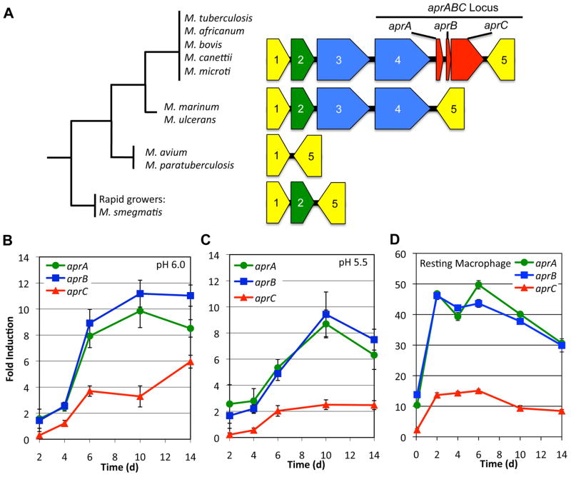
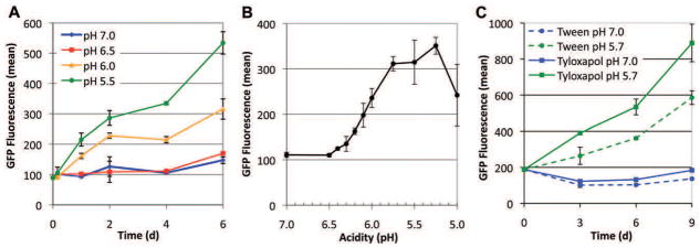
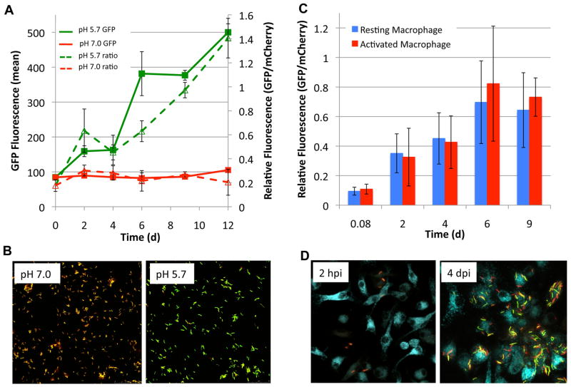
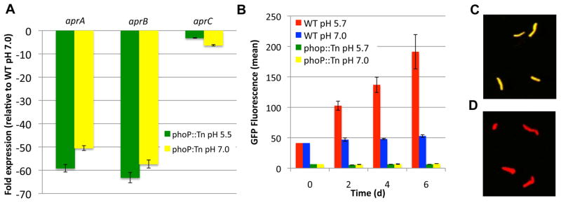
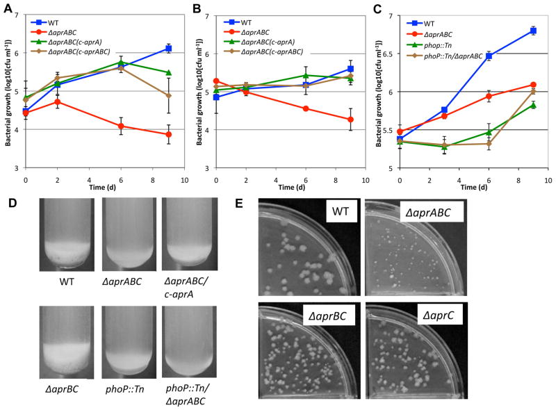
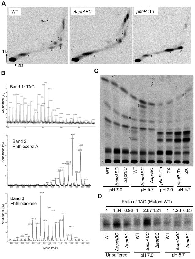
Following phagocytosis by macrophages, Mycobacterium tuberculosis (Mtb) senses the intracellular environment and remodels its gene expression for growth in the phagosome. We have identified an acid and phagosome regulated (aprABC) locus that is unique to the Mtb complex and whose gene expression is induced during growth in acidic environments in vitro and in macrophages. Using the aprA promoter, we generated a strain that exhibits high levels of inducible fluorescence in response to growth in acidic medium in vitro and in macrophages. aprABC expression is dependent on the two-component regulator phoPR, linking phoPR signalling to pH sensing. Deletion of the aprABC locus causes defects in gene expression that impact aggregation, intracellular growth, and the relative levels of storage and cell wall lipids. We propose a model where phoPR senses the acidic pH of the phagosome and induces aprABC expression to fine-tune processes unique for intracellular adaptation of Mtb complex bacteria.


Figures

Figure 1
Figure 1. The aprABC locus is specific to the Mtb complex and exhibits sustained induction during growth in acidic medium in vitro and in MØ
A. Schematic of genes in the region surrounding the aprABC locus. 1=Rv2392, 2=Rv2393, 3=Rv2394, 4=Rv2395, 5=Rv2397. Genes 1 and 5 are involved in sulfur metabolism and are conserved across Mycobacterium species. The phylogenetic tree is an approximate representation of the relationships between the species. The gene colors signify the conservation of genes in a particular species. For example, genes colored red are specific to the Mtb complex species, while genes colored blue are conserved in the Mtb complex as well as M. marinum and M. ulcerans. B-C. Semi-quantitative real time PCR shows in vitro induction of the aprABC locus at pH 6.0 (B) and pH 5.5 (C) when compared to expression at pH 7.0. Error bars represent standard deviation of 4 technical replicates. D. The aprABC locus is induced in resting mouse MØ when compared to the 2h MØ medium control. Expression of aprABC locus genes is slightly repressed during the 2h incubation in MØ medium contributing to the higher magnitude of aprABC locus expression in the MØ as compared to in vitro. The error bars represent the standard deviation of 3 technical replicates.
Figure 2
Figure 2. Acid inducible fluorescence of the CDC1551(aprA′::GFP) reporter
A. The reporter strain was grown in medium buffered at pH 7.0, 6.5, 6.0 or 5.5. Inducible GFP fluorescence is observed at pH 6.0 and 5.5. B. Reporter fluorescence at 6 days post induction in buffered pH medium between 6.5 and 5.0 reveals that CDC1551 (aprA′::GFP) begins to induce GFP fluorescence at pH 6.3 and has an optimal fluorescence between pH 5.75 and 5.25. These pH optima appear tuned to the pH spectrum encountered in the macrophage phagosome. C. Reporter fluorescence is induced at pH 5.7 in media containing either Tween-80 (green circle) or Tyloxapol (green square) detergents, revealing that induction of reporter fluorescence is independent of Tween-80. Fluorescence was measured by counting 25000 cells by flow cytometry. Error bars represent the standard deviation of three biological replicates.
Figure 3
Figure 3. The CDC1551 (aprA′::GFP, smyc′::mCherry) reporter exhibits induced relative fluorescence in response to acid in vitro and during growth in MØ
A. Inducible GFP and relative fluorescence in the Mtb dual fluorescent protein reporter strain. The solid lines show inducible GFP fluorescence at pH 5.7 (green square) and stable GFP fluorescence at pH 7.0 (red square) as determined by the flow cytometry-based counting of 25000 bacteria per sample. Error bars represent the average of 3 biological replicates. The dotted lines show ratiometric measurement of relative fluorescence induction. Induction of relative fluorescence was observed at pH 5.7 (green triangle) with stable relative fluorescence at pH 7.0 (red triangle). B. Micrographs showing increase in GFP fluorescence at pH 5.7 compared with pH 7.0. C. Induction of relative fluorescence in resting and activated mouse MØ. Relative fluorescence was quantified by confocal microscopy by measuring GFP and mCherry fluorescence of ~100 individual intracellular bacteria. Error bars represent the standard deviation. D. Confocal micrographs showing the induction of GFP fluorescence in activated MØ 4 d post infection (dpi) as compared to 2 h post infection (hpi). MØ were loaded prior to infection with AlexaFluor647 labeled 10000 MW dextran (false colored cyan) which is trafficked to the lysosomes. Images presented are maximum projections of a 10 micron Z-stack.
Figure 4
Figure 4. Expression of the aprABC locus is dependent on phoP
A. The aprABC locus is expressed at low levels in the phoP::Tn mutant. qRT-PCR comparing aprA, aprB and aprC expression in the WT at pH 7.0 and the phoP::Tn mutant at pH 7.0 (yellow) and 5.5 (green) 2 h post acid stress. Error bars represent the standard deviation of 3 technical replicates. B. Comparison of acid inducible GFP fluorescence in the CDC1551 and phoP::Tn (aprA′::GFP, smyc′::mCherry) reporter strains. GFP fluorescence was measured by flow cytometry counting of 25000 bacteria per replicate. Error bars are the standard deviation of 3 biological replicates. C-D. Induction of GFP fluorescence in WT reporter (C) but not in the phoP::Tn mutant (D).
Figure 5
Figure 5. Deletion of the aprABC locus causes a defect in intracellular growth and cellular aggregation
A and B. Survival assays comparing WT, ΔaprABC mutant and complemented strains with aprA alone (c-aprA) or the whole aprABC locus (c-aprABC) in resting (A) and activated (B) mouse MØ. Error bars represent standard deviation of three technical replicates. Lower growth of the ΔaprABC mutant compared with the WT was observed in 4 independent biological replicates, although the magnitude of the defect varies between ~1 to ~2 logs of differential growth, depending on the assay (as demonstrated in panel C). C. Survival assay of WT, ΔaprABC mutant, phoP::Tn mutant and the phoP::Tn/ΔaprABC double mutant in resting mouse MØ. D. Aggregation and sedimentation of cultures grown in 7H9 in the absence of Tween-80. E. The ΔaprABC mutant grows as small colonies on agar plates. Strains expressing aprA (e.g. WT, ΔaprBC and ΔaprC) grow as normal colonies. Complementation with aprA elsewhere on the chromosome also returns colonies to normal size (Figure S5).
Figure 6
Figure 6. The deletion of the aprABC locus alters the accumulation of mycobacterial lipids
A. 2D TLC of apolar cell associated Mtb lipids extracted from Mtb grown at pH 5.7. Equal counts (10000 cpm) of 14C-acetate labeled lipids are separated by TLC. The ΔaprABC and phoP::Tn mutants exhibit altered patterns of lipid accumulation. Spots with altered accumulation are numbered 1–6, with the numbers corresponding to the same spots on each TLC. B. Identification of lipid species by MALDI-TOF mass spectrometry. Spectra of [M + Na]+ ions of three identified species: Band 1: Triacylglycerols, Band 2: Phthiocerol A dimycocerate, Band 3: Phthiodiolone dimycocerate. C. Separation of 14C labeled apolar lipids in 1D (petroleum ether:ethyl acetate 98:2). Notice that band 6 only accumulates in the aprABC mutant at pH 5.7, in the absence of aprA and presence of phoP. Phthiocerol A and Phthiodiolone mycocerates accumulate at higher levels in the phoP::Tn mutants and lower levels in the ΔaprABC and ΔaprBC mutants. 2X= phopP:Tn/ΔaprABC double mutant. D. Increased accumulation of TAGs specifically in the ΔaprABC mutant, in an aprA-dependent manner. The presence of aprA, either in the ΔaprBC mutant or complemented elsewhere in the genome (Figure S7A), returns TAG accumulation to WT levels.
Figure 7
Figure 7. Deletion of the aprABC locus causes global changes to mycobacterial gene expression levels with specific dependence on aprA and aprC
A. The gene expression scatter plot represents a direct comparison between the ΔaprABC mutant and WT. All presented genes have a p-value <0.05 and cutoff lines are presented at 1.4X. The vertical axis presents relative expression of genes in the ΔaprABC mutant compared to the WT, where genes that are “up” are higher in the mutant and “down” are lower in the mutant. Highlighted genes are induced (red) or repressed (blue) in an aprA-dependent manner and listed in Table 1. B and C. Expression profiles of aprA dependent genes from panel A in all four mutants, including profiles where differential expression in ΔaprABC and phoP::Tn mutants are similar (B) or different (C). D. Scatter plot of ΔaprABC differentially regulated genes (same as panel A), with aprC dependent genes highlighted. Genes that are induced (red) or repressed (blue) in an aprC-dependent manner and listed in Table S1 and S2. E and F. Expression profiles of aprC-dependent genes from panel D in all four mutants, including profiles where differential expression in ΔaprABC and phoP::Tn mutants are similar (E) or different (F).

Similar articles

Cited by

References

    1. Asensio JG, Maia C, Ferrer NL, Barilone N, Laval F, Soto CY, et al. The virulence-associated two component PhoP-PhoR system controls the biosynthesis of polyketide-derived lipids in Mycobacterium tuberculosis. J Biol Chem. 2006;281:1313–1316. - PubMed
    1. Basu S, Pathak SK, Banerjee A, Pathak S, Bhattacharyya A, Yang ZH, et al. Execution of macrophage apoptosis by PE_PGRS33 of Mycobacterium tuberculosis is mediated by toll-like receptor 2-dependent release of tumor necrosis factor-alpha. J Biol Chem. 2007;282:1039–1050. - PubMed
    1. Brennan MJ, Delogu G, Chen YP, Bardarov S, Kriakov J, Alavi M, Jacobs WR. Evidence that mycobacterial PE_PGRS proteins are cell surface constituents that influence interactions with other cells. Infect Immun. 2001;69:7326–7333. - PMC - PubMed
    1. Carroll P, Schreuder LJ, Muwanguzi-Karugaba J, Wiles S, Robertson BD, Ripoll J, et al. Sensitive Detection of Gene Expression in Mycobacteria under Replicating and Non-Replicating Conditions Using Optimized Far-Red Reporters. Plos One. 2010;5:e9823. - PMC - PubMed
    1. Chang JC, Miner MD, Pandey AK, Gill WP, Harik NS, Sassetti CM, Sherman DR. igr Genes and Mycobacterium tuberculosis Cholesterol Metabolism. J Bacteriol. 2009;191:5232–5239. - PMC - PubMed

Publication types

MeSH terms