. 2011 Mar 14:9:5.
doi: 10.1186/1479-0556-9-5.

B cells Can Modulate the CD8 Memory T Cell after DNA Vaccination Against Experimental Tuberculosis

Affiliations

B cells Can Modulate the CD8 Memory T Cell after DNA Vaccination Against Experimental Tuberculosis

Luciana P Almeida et al. Genet Vaccines Ther. .

Abstract

Background: Although B cells are important as antigen presenting cells (APC) during the immune response, their role in DNA vaccination models is unknown.

Methods: In this study in vitro and in vivo experiments were performed to evaluate the ability of B cells to protect mice against Mycobacterium tuberculosis challenge.

Results: In vitro and in vivo studies showed that B cells efficiently present antigens after naked plasmid pcDNA3 encoding M. leprae 65-kDa heat shock protein (pcDNA3-Hsp65) internalization and protect B knock-out (BKO) mice against Mycobacterium tuberculosis infection. pcDNA3-Hsp65-transfected B cells adoptively transferred into BKO mice rescued the memory phenotypes and reduced the number of CFU compared to wild-type mice.

Conclusions: These data not only suggest that B cells play an important role in the induction of CD8 T cells but also that they improve bacterial clearance in DNA vaccine model.


Figures

Figure 1
Figure 1
Proliferation of CD4 and CD8 T lymphocytes obtained after pcDNA3-HSP65 immunization. (A-B) C57BL/6 wild-type (WT) mice were immunized three times with 100 μg of naked pcDNA3 encoding M. leprae 65-kDa heat shock protein (pcDNA3-HSP65)/mouse in fifteen days interval. Fifteen (A) or thirty days (B) after the last immunization, the spleens were cultured in the presence of peritoneal macrophages and 50 μg of recombinant Hsp5 for three days to re-stimulate Hsp65 specific cells. CD8 and CD4 T cells were sorted and incubated for three days with different antigen presenting cells that had been previously electroporated with pcDNA3, pcDNA3-Hsp65 or a mock control (medium). The proliferation was measured by CFSE staining dilution by flow cytometry and the mock control values were subtracted from the mock vector and pcDNA3-Hsp65 ones. Assays were performed in triplicate and the results represent the mean ± SD of at least two independent experiments. *P < 0,05 versus other stimuli (One-way ANOVA with Tukey's post-test). (C-D) Gene profile of GATA3, T-bet and Foxp3 from total RNA of both population were evaluated by real-time PCR 15 (C) and 30 (D) days after the last dose of the vaccine and incubated for three days with B cells as previously described. The level of expression was calculated using the mock cell as an internal control. The data is representative of 2 independent experiments.
Figure 2
Figure 2
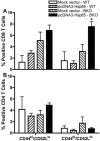
Phenotypical analysis of memory cells in the spleen of reconstituted BKO mice. 1 × 10⁶ electroporated B lymphocytes (pcDNA3-Hsp65 or mock vector) were injected by endovenous route (orbital plexus) in WT and B cell deficient mice. After seven days, the mice were euthanized for analysis of CD44 and CD62L on CD8 (A) and CD4 (B) T cells, by flow cytometry. Assays were performed in triplicate and the results represent the mean ± SD of at least two independent experiments. *P < 0,05 versus wild-type mice (One-way ANOVA with Tukey's post-test).
Figure 3
Figure 3
Colony forming units (CFU) of M. tuberculosis in the lung of infected mice. The BKO mice which received electroporated B cells and control wild-type mice, were inoculated with 1x10⁵ M. tuberculosis. Thirty days post infection, the CFU in the lung homogenates were analyzed. Assays were performed in triplicate and the results represent the mean ± SD of at least two independent experiments. *P < 0,05 versus wild-type mice (One-way ANOVA with Tukey's post-test).

References

    1. Gurunathan S, Klinman DM, Seder RA. DNA vaccines: immunology, application, and optimization*. Annu Rev Immunol. 2000;18:927–974. doi: 10.1146/annurev.immunol.18.1.927. - DOI - PubMed
    1. Tudor D, Dubuquoy C, Gaboriau V, Lefevre F, Charley B, Riffault S. TLR9 pathway is involved in adjuvant effects of plasmid DNA-based vaccines. Vaccine. 2005;23:1258–1264. doi: 10.1016/j.vaccine.2004.09.001. - DOI - PubMed
    1. Bonato VL, Lima VM, Tascon RE, Lowrie DB, Silva CL. Identification and characterization of protective T cells in hsp65 DNA-vaccinated and Mycobacterium tuberculosis-infected mice. Infect Immun. 1998;66:169–175. - PMC - PubMed
    1. Lowrie DB, Tascon RE, Bonato VL, Lima VM, Faccioli LH, Stavropoulos E, Colston MJ, Hewinson RG, Moelling K, Silva CL. Therapy of tuberculosis in mice by DNA vaccination. Nature. 1999;400:269–271. doi: 10.1038/22326. - DOI - PubMed
    1. Coelho-Castelo AA, Santos RR Junior, Bonato VL, Jamur MC, Oliver C, Silva CL. B-lymphocytes in bone marrow or lymph nodes can take up plasmid DNA after intramuscular delivery. Hum Gene Ther. 2003;14:1279–1285. doi: 10.1089/104303403767740812. - DOI - PubMed