. 2011 May 15;509(2):147-56.
doi: 10.1016/j.abb.2011.03.004. Epub 2011 Mar 21.

sFRP-1 binds via its netrin-related motif to the N-module of thrombospondin-1 and blocks thrombospondin-1 stimulation of MDA-MB-231 breast carcinoma cell adhesion and migration

Affiliations

sFRP-1 binds via its netrin-related motif to the N-module of thrombospondin-1 and blocks thrombospondin-1 stimulation of MDA-MB-231 breast carcinoma cell adhesion and migration

Gema Martin-Manso et al. Arch Biochem Biophys. .

Abstract

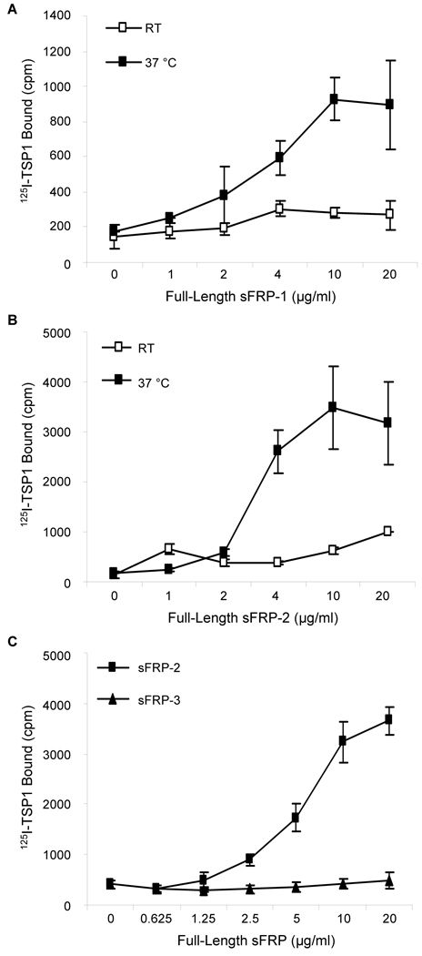
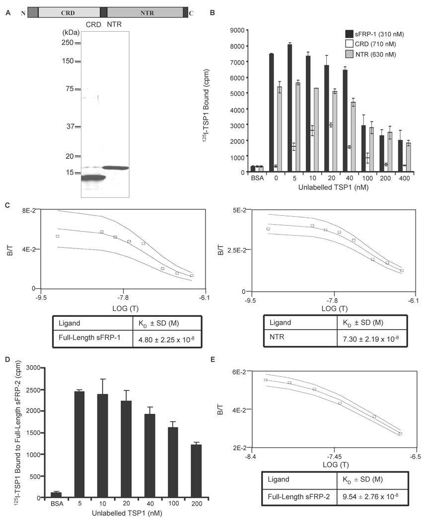
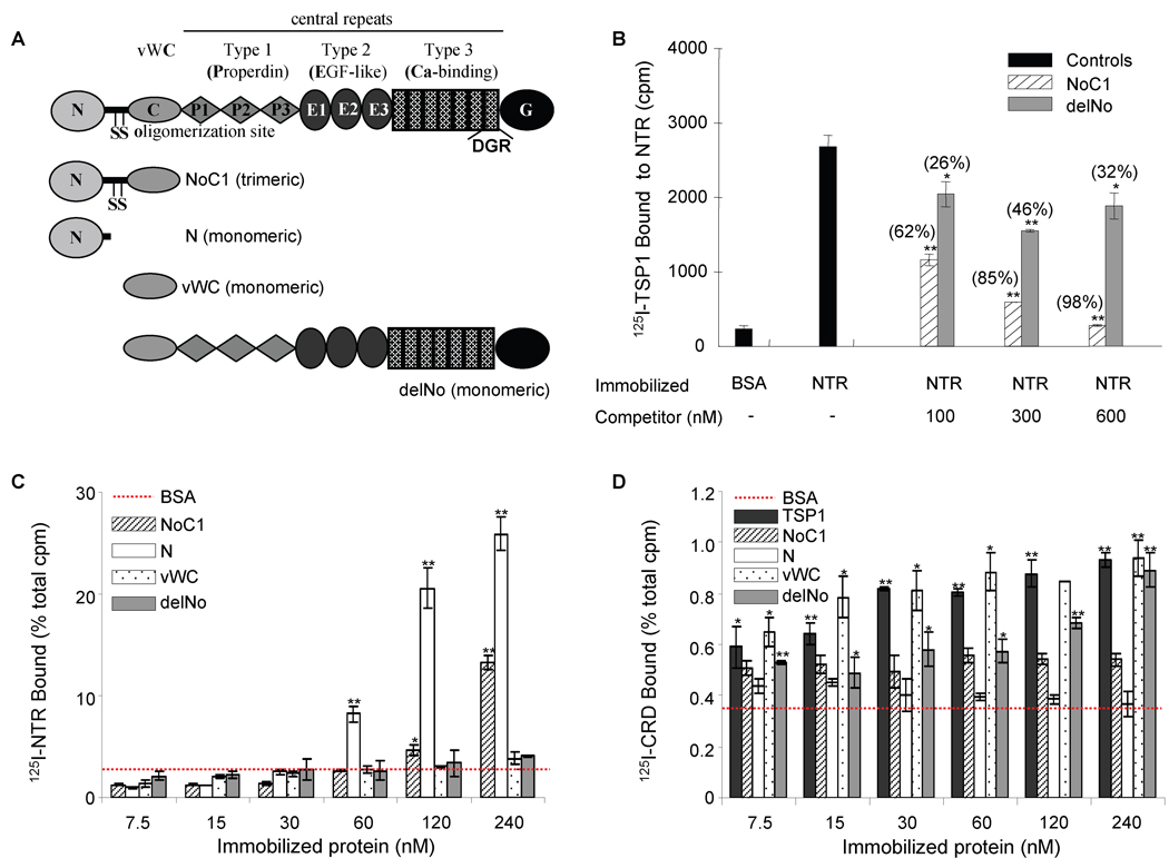
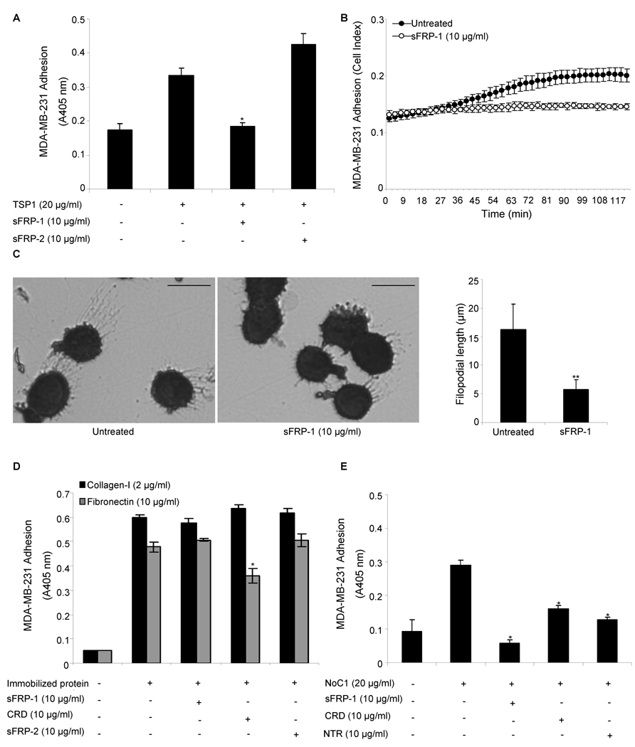
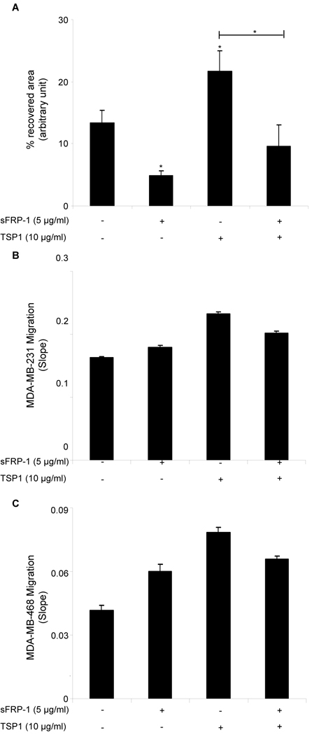
Secreted frizzled-related protein (sFRP)-1 is a Wnt antagonist that inhibits breast carcinoma cell motility, whereas the secreted glycoprotein thrombospondin-1 stimulates adhesion and motility of the same cells. We examined whether thrombospondin-1 and sFRP-1 interact directly or indirectly to modulate cell behavior. Thrombospondin-1 bound sFRP-1 with an apparent K(d)=48nM and the related sFRP-2 with a K(d)=95nM. Thrombospondin-1 did not bind to the more distantly related sFRP-3. The association of thrombospondin-1 and sFRP-1 is primarily mediated by the amino-terminal N-module of thrombospondin-1 and the netrin domain of sFRP-1. sFRP-1 inhibited α3β1 integrin-mediated adhesion of MDA-MB-231 breast carcinoma cells to a surface coated with thrombospondin-1 or recombinant N-module, but not adhesion of the cells on immobilized fibronectin or type I collagen. sFRP-1 also inhibited thrombospondin-1-mediated migration of MDA-MB-231 and MDA-MB-468 breast carcinoma cells. Although sFRP-2 binds similarly to thrombospondin-1, it did not inhibit thrombospondin-1-stimulated adhesion. Thus, sFRP-1 binds to thrombospondin-1 and antagonizes stimulatory effects of thrombospondin-1 on breast carcinoma cell adhesion and motility. These results demonstrate that sFRP-1 can modulate breast cancer cell responses by interacting with thrombospondin-1 in addition to its known effects on Wnt signaling.


Figures

Figure 1
Figure 1. Temperature dependence of 125I-TSP1 binding to recombinant sFRP-1 and sFRP-2
Solid phase binding assays were assessed to quantify 125I-TSP1 (0.5 µg/ml, 50 µl/well) binding to full-length recombinant human sFRP-1 (A), sFRP-2 (B) and sFRP-3 (C) coated onto microtiter plate wells. Nonspecific binding sites were blocked with 3% (w/v) BSA in DPBS containing Ca2+ and Mg2+, at room temperature for 1 h. Binding was performed at room temperature or 37°C in a humidified atmosphere at pH 7.4 for 3 h. The results are representative of two independent experiments performed in triplicate.
Figure 2
Figure 2. Equilibrium binding of 125I-TSP1 to sFRP-1 and sFRP-2
Schematic diagram of sFRP-1 modular structure and Coomassie blue-stained SDS-polyacrylamide gel containing purified CRD and NTR domain of sFRP-1 (A). 125I-TSP1 binding to full-length sFRP-1 or the sFRP-1 domains (CRD and NTR domain) was performed in microtiter plate wells coated with 50 µl/well of 10 µg/ml of each protein. Nonspecific binding was blocked with 3% (w/v) BSA in DPBS containing Ca2+ and Mg2+, at room temperature for 1 h. Binding was measured at 37°C in the presence of competing concentrations of unlabelled TSP1 (B). The Ligand program was used for quantitative analysis of the binding data. Results for sFRP-1 and NTR domain (C left and right, respectively) are presented as displacement plots, and are representative of three (sFRP-1) and two (NTR domain) independent experiments performed in triplicate. Microtiter plate wells were coated using 50 µl/well of 5 µg/ml of full-length sFRP-2. Nonspecific binding was blocked with 3% (w/v) BSA in DPBS containing Ca2+ and Mg2+, at room temperature for 1 h. 125I-TSP1 binding was measured at 37°C in the presence of the indicated concentrations of unlabelled TSP1 (D). The Ligand program was used for quantitative analysis of competitive binding. Results are presented as displacement plots (E), and are representative of two independent experiments performed in triplicate.
Figure 3
Figure 3. Localization of the sFRP-1 binding site in TSP1
20 µg/ml of recombinant NTR domain were absorbed onto microtiter plate wells (B), and 125I-TSP1 binding was measured at 37°C in the presence of competing concentrations of the indicated unlabelled recombinant regions of TSP1 (as shown in A). Nonspecific sites were blocked with 3% (w/v) BSA in DPBS containing Ca2+ and Mg2+. Data were normalized and presented in brackets as percent of inhibition. The results are representative of three independent experiments, each performed in triplicate. 125I-NTR domain (C) and 125I-CRD (D) binding to the indicated concentrations of TSP1 or TSP1 recombinant regions coated onto microtiter wells was determined as above. Data were normalized and are presented as percent of total cpm. The results are representative of four (125I-NTR) and two (125I-CRD) independent experiments, each performed in triplicate.
Figure 4
Figure 4. sFRP modulation of breast cancer cell adhesion on TSP1
MDA-MB-231 breast cancer cell adhesion on immobilized TSP1 (20 µg/ml) (A–C), type-I collagen (2 µg/ml) or fibronectin (10 µg/ml) (D), NoC1 (20 µg/ml) (E) was assessed in the absence or presence of 10 µg/ml of full-length recombinant sFRP-1, CRD and NTR domain of sFRP-1, or sFRP-2. After 60 min incubation, cell adhesion was quantified by colorimetric detection of cell-associated hexosaminidase activity (A, D–E), using the RT-CES system (ACEA Biosciences) (B) or stained with Diff-Quik and imaged (C; bar = 15 µm). Representative images are shown, and filopodia length (mean ± SD) was quantified using Image J software (right panel) as described in Materials and Methods. The results are representative of three independent experiments, each performed in triplicate.
Figure 5
Figure 5. sFRP-1 inhibits TSP1-mediated breast cancer cell migration
Confluent monolayers of MDA-MB-231 cells were treated with full-length recombinant sFRP-1 (5 µg/ml) and TSP1 (10 µg/ml). After 45 min, monolayers were scratched, and 1 h later the percent of recovered area was calculated on four wound edges per condition. The results are representative of three independent experiments (A). 100 µl cell suspension of 4×10⁵ cells/ml MDA-MB-231 cells (B) or MDA-MB-468 cells (C) were added to the upper chamber of the CIM-Plate 16 in the presence or absence of full-length recombinant sFRP-1 (5 µg/ml) and TSP1 (10 µg/ml). After 30 min incubation, cell migration towards 10% FBS was monitored in real time using an impedance-based system and measurements were automatically collected by the analyzer every 5 min for up to 4 h. The results are presented as slope (changes in cell index/hour). The results are representative of two (B) to three (C) independent experiments performed in duplicate.

References

    1. Baenziger NL, Brodie GN, Majerus PW. Proc Natl Acad Sci U S A. 1971;68:240–243. - PMC - PubMed
    1. Bonnefoy A, Moura R, Hoylaerts MF. Cell Mol Life Sci. 2008;65:713–727. - PMC - PubMed
    1. Isenberg JS, Romeo MJ, Yu C, Yu CK, Nghiem K, Monsale J, Rick ME, Wink DA, Frazier WA, Roberts DD. Blood. 2008;111:613–623. - PMC - PubMed
    1. Good DJ, Polverini PJ, Rastinejad F, Le Beau MM, Lemons RS, Frazier WA, Bouck NP. Proc Natl Acad Sci U S A. 1990;87:6624–6628. - PMC - PubMed
    1. Taraboletti G, Roberts D, Liotta LA, Giavazzi R. J Cell Biol. 1990;111:765–772. - PMC - PubMed

Publication types

MeSH terms

Substances