. 2011 May 10:1389:50-60.
doi: 10.1016/j.brainres.2011.03.016. Epub 2011 Mar 12.

Hypoxia-induced angiogenesis is delayed in aging mouse brain

Affiliations

Hypoxia-induced angiogenesis is delayed in aging mouse brain

Girriso F Benderro et al. Brain Res. .

Abstract

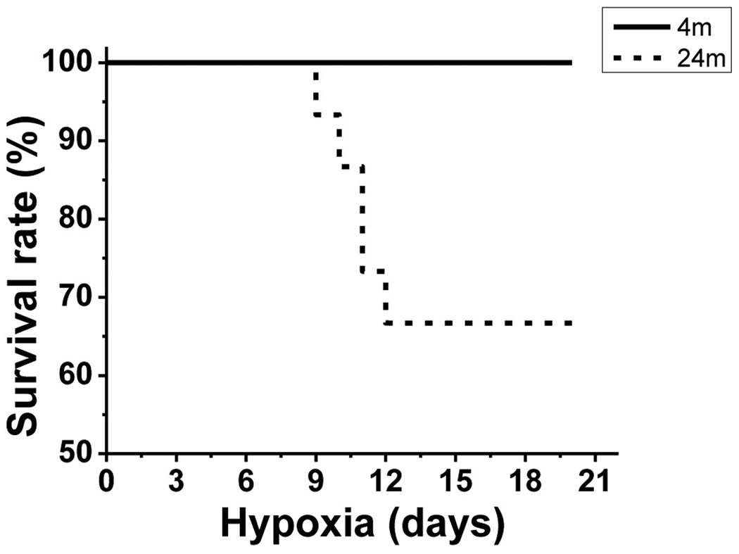
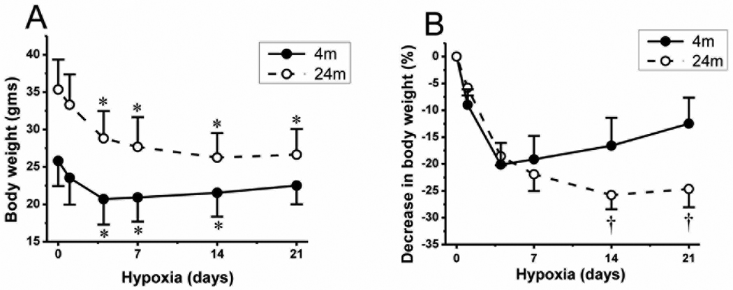
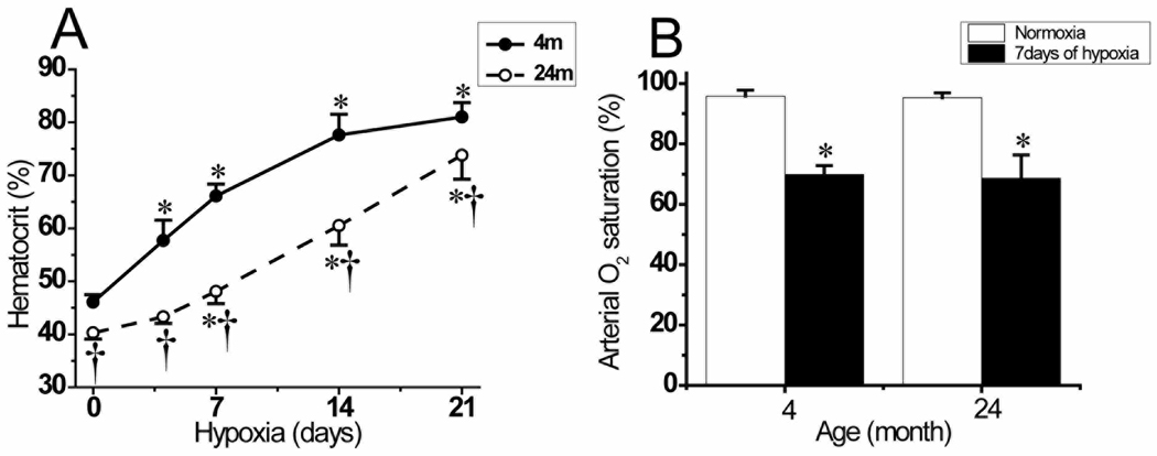
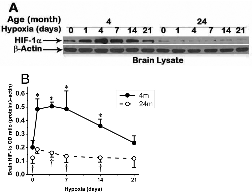
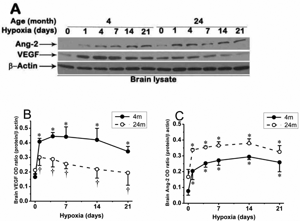
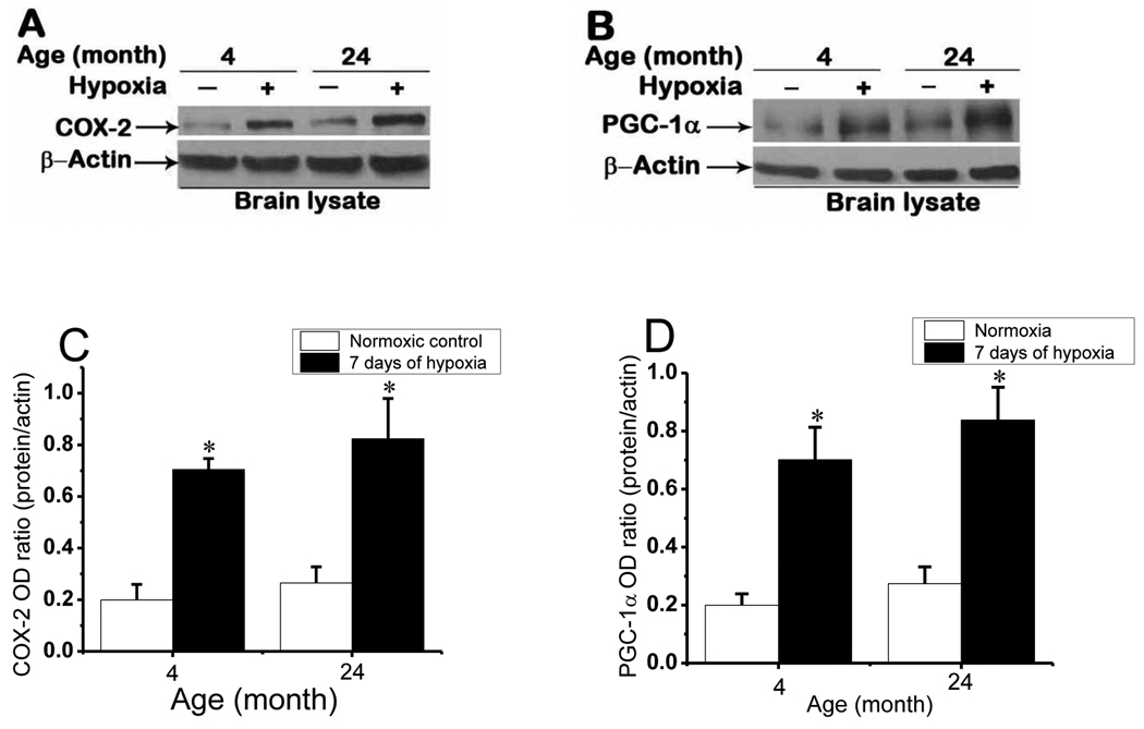
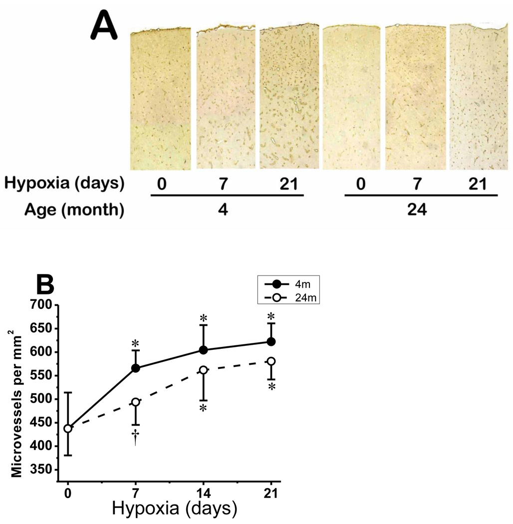
Chronic moderate hypoxia results in systemic and central nervous system adaptations that allow acclimatization. Long-term responses to hypoxia involve systemic physiological changes, metabolic regulation, and vascular remodeling. To investigate whether aging affects systemic and cerebral angiogenic adaptational changes in response to prolonged hypoxia, the present study assessed the responses of 4month old ("young") C57BL/6 mice and 24month old ("aged") C57BL/6 mice to chronic hypobaric hypoxia of 0.4atm (290torr). Compared to young mice, delayed body weight-loss recovery and a lag in polycythemic response were observed in aged mice. As previously shown, hypoxia inducible factor-1α (HIF-1α) accumulation was attenuated and vascular endothelial growth factor (VEGF) expression was decreased in the cerebral cortex of aged mice. Conversely, cyclooxygenase-2 (COX-2), angiopoietin-2 (Ang-2), and peroxisome proliferator-activated receptor gamma coactivator 1-α (PGC-1α) protein upregulation were not affected in the aged mice. Despite an initial delay in cerebral angiogenic response in aged mice in the first week of hypoxia, no significant differences were observed in microvascular density between young and aged mice in normoxia and at 2 and 3weeks of hypoxia. Taken together, these observations indicate that, even though the HIF-1 response to hypoxia is greatly attenuated, HIF-1 independent compensatory pathways are eventually able to maintain baseline and cerebral angiogenic adaptational changes to chronic hypoxia in aged mice. The delayed adaptive response, however, may result in decreased survival in the aged cohort.


Figures

Fig.1
Fig.1
Survival rate. Survival rate in 4 month and 24 month old mice exposed chronic hypoxia, showing 16 of 16 (100%) survival in young mice in 3 weeks of hypoxia, whereas only 10 of 15 (66.7%) of aged mice survived similar hypoxic condition of 3 weeks.
Fig.2
Fig.2
Change in body weight during chronic hypoxia. A: change in gross body weight during 21 days of hypoxia in 4 month old (4m) mice (n=16) and 24 month old (24m) mice (n=10). Error bars are indicated at normoxia (0), and 1, 4, 7, 14, and 21 days of hypoxia. B: relative % decrease in body weight in 4m and 24m old mice during hypoxic exposure. Values are means ± standard deviation (SD). *p < 0.05 compared with corresponding age normoxic control value. p < 0.05 compared with corresponding duration 4 month old mice values.
Fig.3
Fig.3
Hematocrit and arterial oxygen saturation during chronic hypoxia. A: hematocrit values in 4 month old mice under normoxic control (0) (n=19), and 4 (n=10), 7 (n=10), 14 (n=10), and 21 (n=16) days of hypoxia; and 24 month old mice under normoxic control (0) (n=11), and 4 (n=10), 7 (n=11), 14 (n=10), and 21 (n=10) days of hypoxia. B: arterial oxygen saturation in 4month and 24 month old mice in normoxic and 7 days hypoxia exposed mice (n=6 mice in each groups). Values are means ± standard deviation (SD). *p < 0.05 compared with corresponding age normoxic control value. p < 0.05 compared with corresponding duration 4 month old mice values.
Fig. 4
Fig. 4
HIF-1α accumulation in cerebral cortex of mouse in chronic hypoxia. A: attenuated HIF-1α accumulation in aged mice in normoxia and chronic hypoxia compared to the young mice. B: Optical density ratio of HIF-1α normalized to β-actin in normoxia (0), and 1, 4, 7, 14, and 21 days of hypoxia. *p < 0.05 compared with corresponding age normoxic control. p < 0.05 compared with corresponding 4 month old mice values. Each value represents the mean ± SD from 5 mice.
Fig. 5
Fig. 5
Expression of VEGF and Ang-2 in cerebral cortex of 4 month and 24 month old mice. A: VEGF and Ang-2 protein expression in normoxic control and hypoxia in young and aged mice. B: VEGF protein expression optical density ratio in young and aged mice in normoxia (0), and 1, 4, 7, 14, and 21 days of hypoxia and C: Ang-2 protein expression optical density ratio in young and aged mice in normoxia (0), and 1, 4, 7, 14, and 21 days of hypoxia. *p < 0.05 compared with normoxic control values of each category. p < 0.05 aged mice vs corresponding young mice values. Values are mean ± SD, n = 5 mice per time point.
Fig. 6
Fig. 6
COX-2 and PGC-1α expression in 4 and 24 month old mice cerebral cortex in normoxia and 7 days of chronic hypoxia. Representative Western blot analysis of Cox-2 (A) and PGC-1α (B) during normoxic control and 7 days of hypoxic exposure in the young and aged mice. C and D: Optical density ratios of COX-2 and PGC-1α respectively, normalized to β-actin in normoxia and 7 days of hypoxia as a function of age. *p < 0.05 compared with normoxic control values of each age group. Values are mean ± SD, n = 5 mice in each category.
Fig. 7
Fig. 7
Microvascular density in young and aged mice cerebral cortex in normoxic control and prolonged hypoxia. A: composite photomicrograph of GLUT-1–stained sections spanning part of the parietal cortex of 4 and 24 month old mice at normoxia (0), and 7 and 21 days of hypoxia. B: Capillary density count (number per mm2) of GLUT-1-stained sections of 4 month old and 24 month old mice during normoxia (0) and 7, 14 and 21 days of hypoxia. *p < 0.05 compared with corresponding normoxic control. p < 0.05 compared with corresponding 4 month old mice value. Values are mean ± SD, n = 5 mice per time point in each group.

References

    1. Anderson J, Sandhir R, Hamilton ES, Berman NE. Impaired expression of neuroprotective molecules in the HIF-1alpha pathway following traumatic brain injury in aged mice. J. Neurotrauma. 1566;26:1557–1566. - PMC - PubMed
    1. Arany Z, Foo SY, Ma Y, Ruas JL, Bommi-Reddy A, Girnun G, Cooper M, Laznik D, Chinsomboon J, Rangwala SM, Baek KH, Rosenzweig A, Spiegelman BM. HIF-independent regulation of VEGF and angiogenesis by the transcriptional coactivator PGC-1alpha. Nature. 2008;451:1008–1012. - PubMed
    1. Bates DO, Curry FE. Vascular endothelial growth factor increases microvascular permeability via a Ca(2+)-dependent pathway. Am. J. Physiol. 1997;273:H687–H694. - PubMed
    1. Benelli R, Lorusso G, Albini A, Noonan DM. Cytokines and chemokines as regulators of angiogenesis in health and disease. Curr. Pharm. Des. 2006;12:3101–3115. - PubMed
    1. Boero JA, Ascher J, Arregui A, Rovainen C, Woolsey TA. Increased brain capillaries in chronic hypoxia. J. Appl. Physiol. 1999;86:1211–1219. - PubMed

Publication types

MeSH terms

Substances