. 2011 Aug 3;104(2):257-65.
doi: 10.1016/j.physbeh.2011.03.006. Epub 2011 Mar 21.

Immune challenge activates neural inputs to the ventrolateral bed nucleus of the stria terminalis

Affiliations

Immune challenge activates neural inputs to the ventrolateral bed nucleus of the stria terminalis

Michael S Bienkowski et al. Physiol Behav. .

Abstract

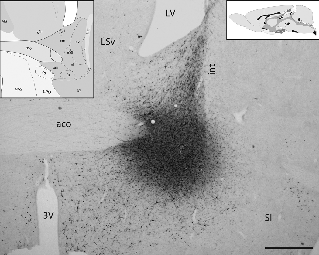
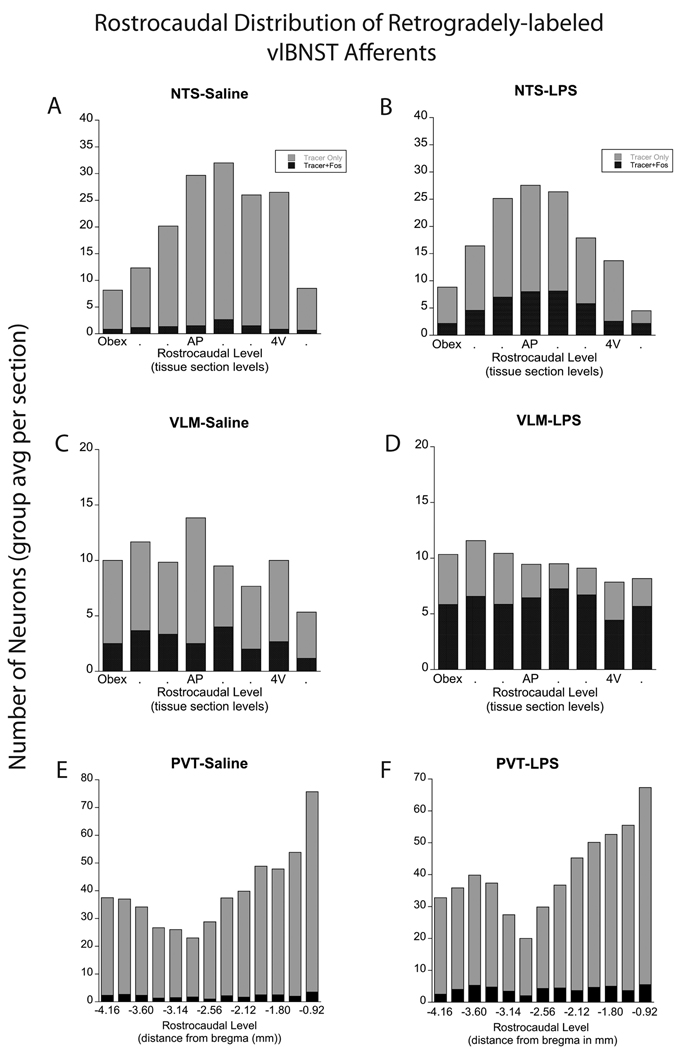
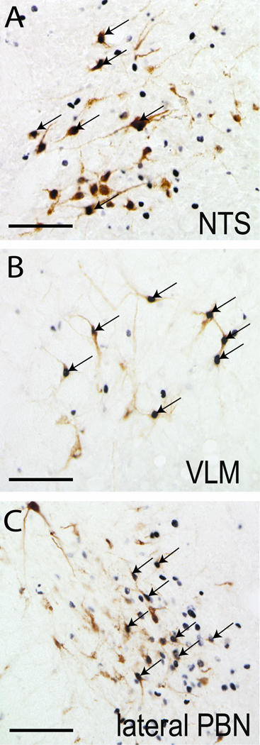
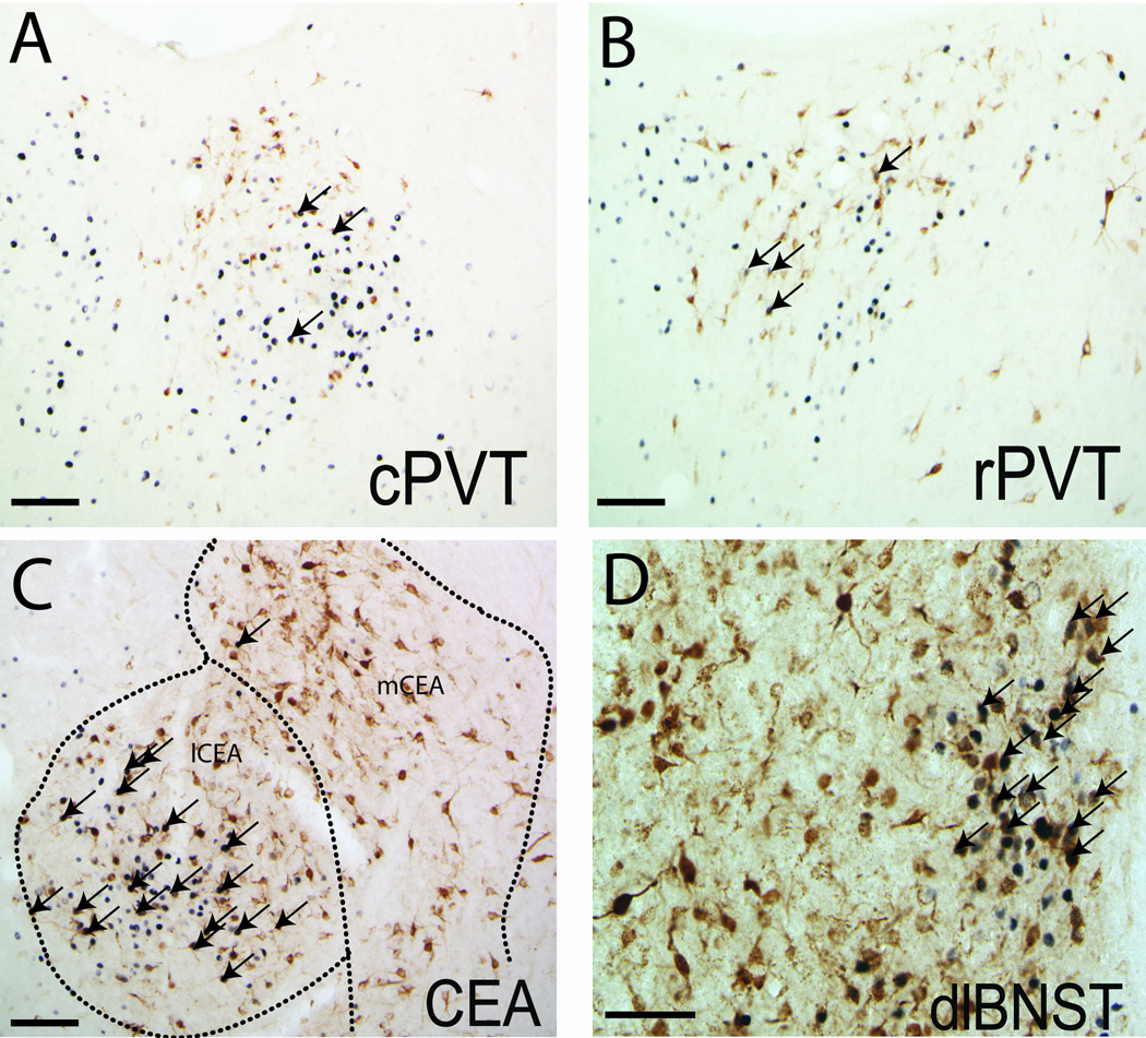
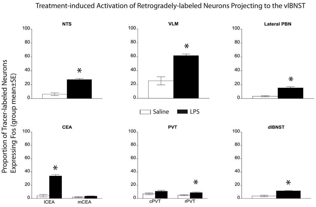
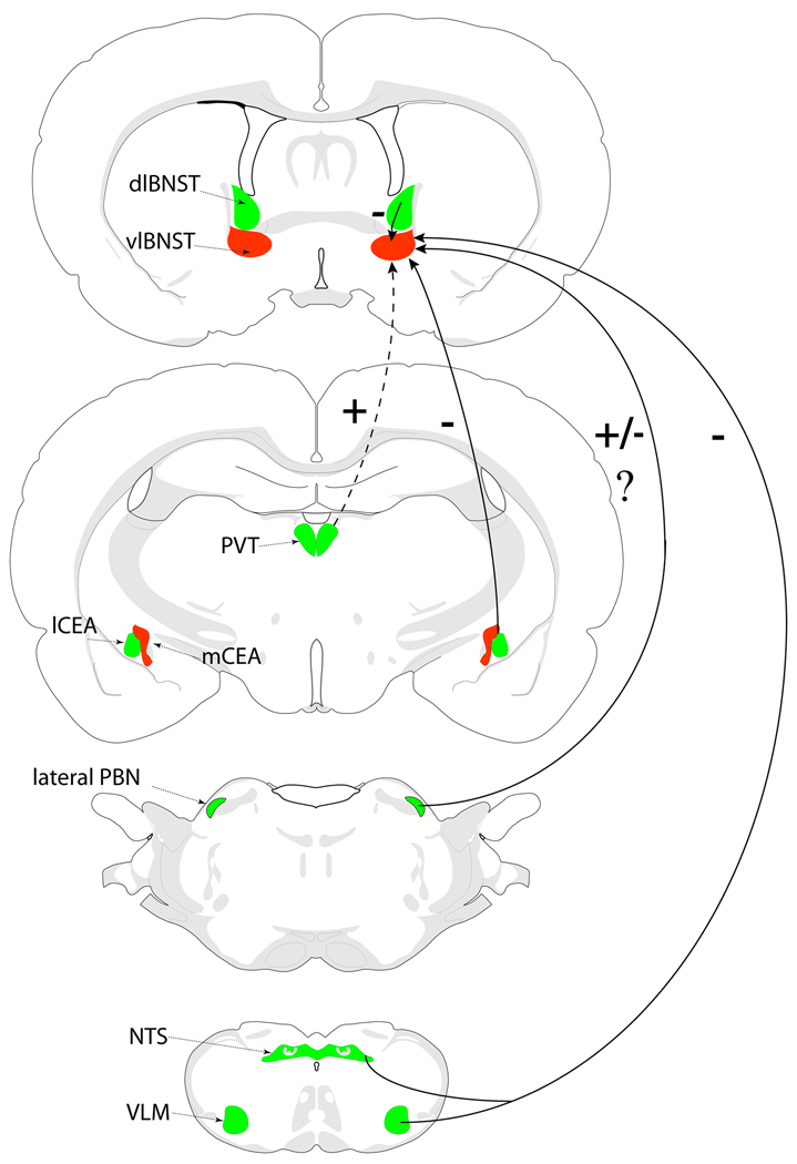
Hypothalamo-pituitary-adrenal (HPA) axis activation in response to infection is an important mechanism by which the nervous system can suppress inflammation. HPA output is controlled by the hypothalamic paraventricular nucleus (PVN). Previously, we determined that noradrenergic inputs to the PVN contribute to, but do not entirely account for, the ability of bacterial endotoxin (i.e., lipopolysacharide, LPS) to activate the HPA axis. The present study investigated LPS-induced recruitment of neural inputs to the ventrolateral bed nucleus of the stria terminalis (vlBNST). GABAergic projections from the vlBNST inhibit PVN neurons at the apex of the HPA axis; thus, we hypothesize that LPS treatment activates inhibitory inputs to the vlBNST to thereby "disinhibit" the PVN and increase HPA output. To test this hypothesis, retrograde neural tracer was iontophoretically delivered into the vlBNST of adult male rats to retrogradely label central sources of axonal input. After one week, rats were injected i.p. with either LPS (200 μg/kg BW) or saline vehicle, and then perfused with fixative 2.5h later. Brains were processed for immunohistochemical localization of retrograde tracer and the immediate-early gene product, Fos (a marker of neural activation). Brain regions that provide inhibitory input to the vlBNST (e.g., caudal nucleus of the solitary tract, central amygdala, dorsolateral BNST) were preferentially activated by LPS, whereas sources of excitatory input (e.g., paraventricular thalamus, medial prefrontal cortex) were not activated or were activated less robustly. These results suggest that LPS treatment recruits central neural systems that actively suppress vlBNST neural activity, thereby removing a potent source of inhibitory control over the HPA axis.


Figures

Figure 1
Figure 1. Retrograde tracer delivery site in the vlBNST
Iontophoretic delivery of FG (shown here) or CTb produced spherical tracer delivery sites. Iontophoretic sites were considered accurate if they were centered within the anterolateral (al) and fusiform (fu) subnuclei of the vlBNST, with minimal spread of the surrounding halo into adjacent regions. Note that labeling observed within the oval subnucleus of the dorsal BNST (ov) and other nearby regions is the product of retrograde transport from the vlBNST tracer delivery site. Atlas figures (insets) from Swanson [43]. The vertical line in the upper right inset indicates the approximate rostrocaudal level depicted in the photomicrograph. 3V=third ventricle, aco= anterior commissure, int=internal capsule, LSv=ventral lateral septum, LV= lateral ventricle, SI=substantia innominata. Scale bar= 1 mm.
Figure 2
Figure 2. Rostrocaudal distribution of retrogradely-labeled neurons in the NTS, VLM, and PVT
Rostrocaudal distribution of retrograde labeling in the NTS, VLM, and PVT in rats after control saline (left panels; n=6) or LPS treatment (right panels; n=8). The number of single- and double-labeled NTS neurons (A, B) was greatest at the rostrocaudal level of the mid area postrema (AP) and just rostral to it. Within the VLM (C, D), single- and double-labeled neurons were distributed more evenly across rostrocaudal levels. The proportion of tracer-labeled NTS and VLM neurons that expressed Fos was significantly increased in rats after LPS treatment (see Fig. 5 and Table 1). Larger numbers of tracer-labeled neurons were present within the rostral half of the PVT (rPVT) compared to the caudal half (cPVT; E, F). LPS treatment produced a small yet significant increase in the number and proportion of double-labeled (activated) neurons in the rPVT, but not in the cPVT, compared to saline control treatment (see Fig. 5 and Table 1). 4V=fourth ventricle.
Figure 3
Figure 3. LPS-induced Fos activation of brainstem vlBNST afferents
LPS treatment activated vlBNST-projecting neurons within the NTS (A), VLM (B), and lateral PBN (C), as evidenced by Fos expression (black nuclear staining) within retrogradely-labeled neurons (brown cytoplasmic labeling). Arrows point out examples of double-labeled neurons. Scale bars = 100 µm.
Figure 4
Figure 4. LPS-induced Fos activation of forebrain vlBNST afferents
Despite the presence of numerous retrogradely-labeled neurons and a relatively high incidence of Fos expression within the caudal (A) and rostral PVT (B), relatively few double-labeled neurons were observed in rats after LPS treatment. Conversely, many retrogradely-labeled neurons in the lCEA (C) and dlBNST (D, particularly prevalent within the oval subnucleus) expressed Fos in rats after LPS treatment. Arrows point out examples of double-labeled neurons. Scale bars = 100 µm.
Figure 5
Figure 5. Proportion of activated vlBNST afferents in LPS vs saline rats
The proportion of tracer-labeled neurons that also were Fos-positive (i.e., double-labeled) was significantly increased within the NTS, VLM, lateral PBN, lCEA, and dlBNST in LPS-treated rats (n=8) compared to saline-injected controls (n=6). Small but still significant increases in activation also were observed within the rPVT in LPS-treated rats vs. controls. Conversely, there was no significant effect of LPS treatment on Fos activation of retrogradely labeled neurons within either the mCEA or the cPVT.
Figure 6
Figure 6. vlBNST inputs converge to promote inhibition of vlBNST activity
Summary diagram of neural inputs to the vlBNST that are recruited/activated (green) or not recruited/activated (red) in response to acute immune challenge with LPS. Together with previous findings, the results of this study suggest that activated neural inputs from the NTS, VLM, lateral PBN, lCEA, and dlBNST converge to promote inhibition of the vlBNST in rats after LPS treatment. Conversely, glutamatergic inputs from the PVT and mPFC (not shown) are weakly activated or not activated in rats after LPS treatment. +, presumably excitatory pathway; −, presumably inhibitory pathway; +/−?, unknown impact. Dashed line, minimal LPS-induced recruitment of this pathway. See text for details.

Similar articles

Cited by

References

    1. Buller KM. Circumventricular organs: gateways to the brain role of circumventricular organs in pro-inflammatory cytokine-induced activation of the hypothalamic- pituitary-adrenal axis. Clinical and Experimental Pharmacology and Physiology. 2001;28:581–589. - PubMed
    1. Ek M, Kurosawa M, Lundeberg T, Ericsson A. Activation of vagal afferents after intravenous injection of interleukin-1beta : role of endogenous prostaglandins. J. Neurosci. 1998;18:9471–9479. - PMC - PubMed
    1. Vitkovic L, Konsman JP, Bockaert J, Dantzer R, Homburger V, Jacque C. Cytokine signals propagate through the brain. Molecular Psychiatry. 2000;5:604–615. - PubMed
    1. Ericsson A, Arias C, Sawchenko PE. Evidence for an intramedullary prostaglandin-dependent mechanism in the activation of stress-related neuroendocrine circuitry by intravenous interleukin-1. J. Neurosci. 1997;17:7166–7179. - PMC - PubMed
    1. Goehler LE, Gaykema RPA. Neural pathways mediating behavioral changes associated with immunological challenge. In: Siegal A, Zalcman S, editors. The Neuroimmunological Basis of Behavior and Mental Disorders. 1st ed. New York: Springer; 2008.

Publication types

Substances