. 2011 Mar 17;9(3):200-211.
doi: 10.1016/j.chom.2011.02.009.

Structural and functional analysis of a plant resistance protein TIR domain reveals interfaces for self-association, signaling, and autoregulation

Affiliations

Structural and functional analysis of a plant resistance protein TIR domain reveals interfaces for self-association, signaling, and autoregulation

Maud Bernoux et al. Cell Host Microbe. .

Abstract

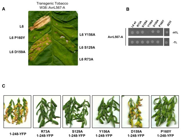
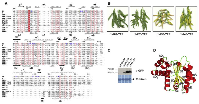
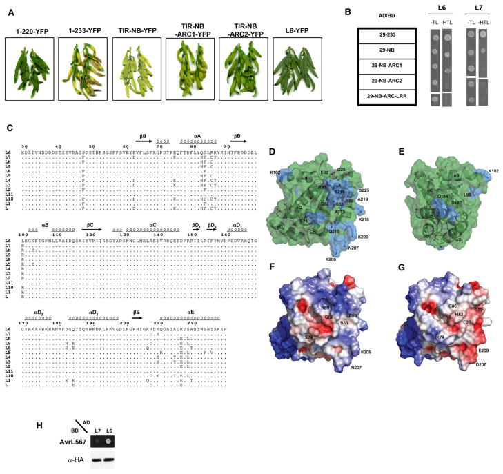
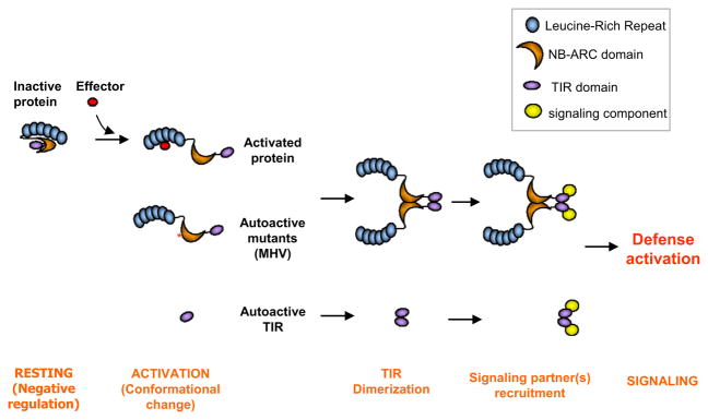
The Toll/interleukin-1 receptor (TIR) domain occurs in animal and plant immune receptors. In the animal Toll-like receptors, homodimerization of the intracellular TIR domain is required for initiation of signaling cascades leading to innate immunity. By contrast, the role of the TIR domain in cytoplasmic nucleotide-binding/leucine-rich repeat (NB-LRR) plant immune resistance proteins is poorly understood. L6 is a TIR-NB-LRR resistance protein from flax (Linum usitatissimum) that confers resistance to the flax rust phytopathogenic fungus (Melampsora lini). We determine the crystal structure of the L6 TIR domain and show that, although dispensable for pathogenic effector protein recognition, the TIR domain alone is both necessary and sufficient for L6 immune signaling. We demonstrate that the L6 TIR domain self-associates, most likely forming a homodimer. Analysis of the structure combined with site-directed mutagenesis suggests that self-association is a requirement for immune signaling and reveals distinct surface regions involved in self-association, signaling, and autoregulation.


Figures

Figure 1
Figure 1. Mutations in L6 TIR Domain Affect HR Induction and Signaling Activation but Not Effector Recognition
(A) Transgenic tobacco W38 expressing AvrL567-A, 4 days after infiltration with A. tumefaciens strains carrying full-length L6 wild-type or TIR domain mutants. (B) Growth of yeast cells coexpressing GAL4-BD::AvrL567-A with GAL4-AD::full-length L6 wild-type, TIR domain mutants or L6 lacking the TIR domain (Δ233). Growth on media lacking tryptophan and leucine (−TL) confirms yeast viability, while growth on media lacking histidine (−HTL) indicates expression of the HIS3 reporter gene due to interaction between the fusion proteins. (C) Flax Hosh plants 12 days after infiltration with A. tumefaciens strains carrying L6 TIR1-248 mutants fused to YFP.
Figure 2
Figure 2. L6 TIR Domain Is Sufficient and Necessary to Trigger Cell Death Signaling
(A) Multiple sequence alignment of TIR domains. Amino acid sequences from the TIR domains of L6 (residues 59–240), N (10–191), RPS4 (15–191), RPP1-WsA (50–229), and RPP1-WsB (83–262) were aligned with the sequences of the TIR domains with known 3D structures: AtTIR (PDB ID 3JRN), PdTIR (3H16), MyD88 (2Z5V), IL-1RAPL (1T3G), TLR1 (1FYV), TLR2 (1FYW), and TLR10 (2J67) using MUSCLE (Edgar 2004). The positions of the secondary structure elements in L6 and TLR2 are shown at the top and bottom, respectively. The alignment was formatted using ESPript (Gouet et al., 2003). Strictly conserved residues are indicated in white letters with a red box and similar residues are indicated in red letters with a red frame, while mutated residues are indicated in bold blue letters. The intron site is indicated by an arrow head. (B) Flax Hosh plants 12 days after infiltration with A. tumefaciens strains carrying L6 TIR domain deletion constructs fused to YFP. (C) Immunoblot detection of L6 TIR-YFP fusions with anti-GFP antibodies 3 days after agroinfiltration in flax leaves. Lower panel shows membrane stained with amido black indicating equal loading of Rubisco. (D) Crystal structure of L6TIR. Ribbon drawing of one of the two molecules in the asymmetric unit. The secondary structure elements and loops are named according to the nomenclature used for TLR1 TIR domain (Xu et al., 2000) and AtTIR (Chan et al., 2010).
Figure 3
Figure 3. L6 TIR Domain Self-Associates
(A) Growth of yeast cells coexpressing GAL4-BD and GAL4-AD L6 TIR domain fusions on synthetic media lacking tryptophan and leucine (−TL) or selective media additionally lacking histidine (−HTL). (B) Immunoblot detection of GAL4-AD and GAL4-BD fusion proteins in yeast. Proteins were detected with anti-HA and anti-Myc antibodies, respectively. (C–F) Solution properties of L6TIR. Red lines indicate the trace from the refractive index detector (arbitrary units) during size exclusion chromatography, and the blue lines are the weight-average molecular weight (Mw; y axis) distribution across the peak determined by MALLS. In (C), the initial concentration of L6TIR is 2 mg/ml and the buffer consists of 10 mM HEPES (pH 7.4) and 150 mM NaCl. (D) Same buffer condition as (C), but the initial protein concentration was 1 mg/ml. (E) Initial protein concentration is 2 mg/ml; buffer contains 0 mM NaCl. (F) Initial protein concentration is 2 mg/ml; buffer contains 500 mM NaCl.
Figure 4
Figure 4. TIR/TIR Domain Interfaces in the L6 TIR Crystal
(A) Ribbon representation of the asymmetric unit interfaces (green and yellow) and the interface related by crystallographic symmetry (yellow and cyan). (B) As in (A), with the residues involved in close contacts (at distances <4 Å ) shown in wireframe. Hydrogen bonds/salt bridges are shown as dotted red lines.
Figure 5
Figure 5. The NB-ARC Domain Regulates TIR Autoactivity and Self-Association
(A) Flax Hosh plants 12 days after infiltration with A. tumefaciens strains carrying truncated L6 constructs fused to YFP. (B) Growth of yeast cells coexpressing GAL4-BD and GAL4-AD fused to truncated L6 and L7 constructs on synthetic media lacking tryptophan and leucine (−TL) or selective media additionally lacking histidine (−HTL). (C) Comparison of allelic variants of the L6 TIR domain. Sequence alignment of the TIR domain region of different L alleles. The positions of polymorphic residues are highlighted. (D and E) Transparent surface representation of L6 TIR, polymorphic regions present in allelic variants are shown in blue. The molecule in E is oriented 135° around the vertical axis compared to (D). (F and G) Surface representations of L6TIR (F) and a homology model of the L7 TIR domain (G), with electrostatic potential (calculated using APBS; Baker et al., 2001) mapped to the surface. Coloring is continuous going from blue (potential +5 kt/e) through white to red (potential −5 kt/e). The molecules are oriented as in (D). (H) Growth of yeast cells coexpressing GAL4-BD::AvrL567-A with GAL4-AD::full-length L7 or L6 on selective media lacking histidine (−HTL). GAL4 AD fusion proteins were detected with anti-HA antibodies.
Figure 6
Figure 6. Proposed Model for Activation of TIR-NB-LRRs Proteins
In the absence of a recognized effector protein, intramolecular interactions keep the protein in a resting conformation where the TIR domain dimerization interface is not exposed. Upon activation, negative regulation is released, probably through nucleotide exchange in the NB domain, and conformational change exposes the TIR domain for homodimerization and the TIR domain signaling interface for interaction with signaling proteins. This induces a signaling cascade leading to innate immunity. Autoactive variants, such as the MHV mutant (contains a D-to-V mutation in the MHD motif in the ARC2 subdomain) or the TIR domain alone, circumvent the need for effector recognition.

Similar articles

Cited by

References

    1. Adams PD, Afonine PV, Bunkóczi G, Chen VB, Davis IW, Echols N, Headd JJ, Hung LW, Kapral GJ, Grosse-Kunstleve RW, et al. PHENIX: a comprehensive Python-based system for macromolecular structure solution. Acta Crystallogr D Biol Crystallogr. 2010;66:213–221. - PMC - PubMed
    1. Ashkenazy H, Erez E, Martz E, Pupko T, Ben-Tal N. ConSurf 2010: calculating evolutionary conservation in sequence and structure of proteins and nucleic acids. Nucleic Acids Res Suppl. 2010;38:W529–W533. - PMC - PubMed
    1. Baker NA, Sept D, Joseph S, Holst MJ, McCammon JA. Electrostatics of nanosystems: application to microtubules and the ribosome. Proc Natl Acad Sci USA. 2001;98:10037–10041. - PMC - PubMed
    1. Bao Q, Shi Y. Apoptosome: a platform for the activation of initiator caspases. Cell Death Differ. 2007;14:56–65. - PubMed
    1. Bernoux M, Timmers T, Jauneau A, Briere C, de Wit PJ, Marco Y, Deslandes L. RD19, an Arabidopsis cysteine protease required for RRS1-R-mediated resistance, is relocalized to the nucleus by the Ralstonia solanacearum PopP2 effector. Plant Cell. 2008;20:2252–2264. - PMC - PubMed

Publication types

MeSH terms

Substances

LinkOut - more resources