. 2011 Apr 1;203(7):1021-30.
doi: 10.1093/infdis/jiq136.

Ultrastructural, immunofluorescence, and RNA evidence support the hypothesis of a "new" virus associated with Kawasaki disease

Affiliations

Ultrastructural, immunofluorescence, and RNA evidence support the hypothesis of a "new" virus associated with Kawasaki disease

Anne H Rowley et al. J Infect Dis. .

Abstract

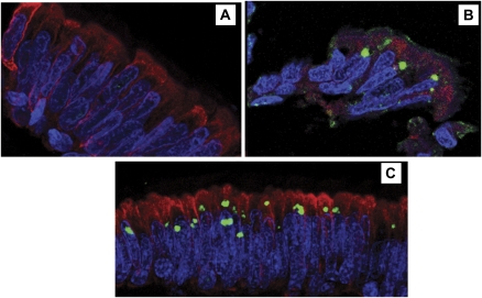
Background: Intracytoplasmic inclusion bodies (ICI) have been identified in ciliated bronchial epithelium of Kawasaki disease (KD) patients using a synthetic antibody derived from acute KD arterial IgA plasma cells; ICI may derive from the KD etiologic agent.

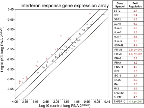
Methods: Acute KD bronchial epithelium was subjected to immunofluorescence for ICI and cytokeratin, high-throughput sequencing, and transmission electron microscopy (TEM). Interferon pathway gene expression profiling was performed on KD lung.

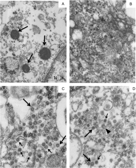
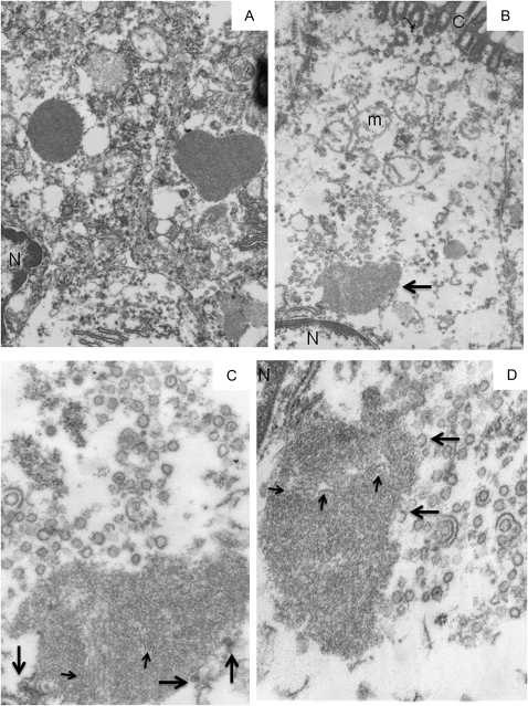
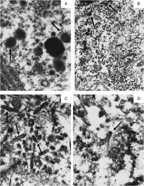
Results: An intermediate filament cytokeratin "cage" was not observed around KD ICI, making it unlikely that ICI are overproduced or misfolded human protein aggregates. Many interferon-stimulated genes were detected in the bronchial epithelium, and significant modulation of the interferon response pathway was observed in the lung tissue of KD patients. No known virus was identified by sequencing. Aggregates of virus-like particles (VLP) were detected by TEM in all 3 acute KD patients from whom nonembedded formalin-fixed lung tissue was available.

Conclusions: KD ICI are most likely virus induced; bronchial cells with ICI contain VLP that share morphologic features among several different RNA viral families. Expedited autopsies and tissue fixation from acute KD fatalities are urgently needed to more clearly ascertain the VLP. These findings are compatible with the hypothesis that the infectious etiologic agent of KD may be a "new" RNA virus.


Figures

Figure 1.
Figure 1.
Confocal immunofluorescence for Kawasaki Disease intracytoplasmic inclusion bodies (green) and cytokeratin (red) in formalin-fixed, paraffin-embedded cilated bronchial epithelium. A, Compressed Z-stack image of bronchial epithelium of control 8, showing cytoplasmic expression of cytokeratin and absence of ICI. B, single plane image of KD bronchial epithelium from Kawasaki disease (KD) patient 2. Intracytoplasmic inclusion bodies (ICI) are roughly spherical and reside just above nuclei; cytokeratin is in the supranuclear cytoplasm but does not form a “cage” around the ICI. C, Compressed Z-stack image of bronchial epithelium of KD patient 3, showing that cytokeratin is expressed in the cytoplasm but does not form a “cage” around the ICI. Some cells have more than 1 ICI and some ICI are somewhat irregular in shape. Nuclei are stained with 4′,6-diamidino-2-phenylindole (blue). Original magnification: x400 for all panels.
Figure 2.
Figure 2.
Real-time polymerase chain reaction analysis of interferon-stimulated genes in acute Kawasaki Disease and control lung. Interferon α, β response polymerase chain reaction array revealed 21 genes upregulated (>2.5-fold) in Kawasaki disease when compared with that of control lung; IFITM1 and IFITM2 were statistically significantly upregulated. 1 gene on the array, TNFSF10, was significantly downregulated and was the only gene downregulated >2.5-fold. Lines indicate boundaries for 2.5-fold up- or downregulation of genes in this pathway. Genes upregulated are indicated in red, and downregulated genes are in green.
Figure 3.
Figure 3.
Transmission electron microscopy of 3 intracytoplasmic inclusion bodies in 1 cell and of virus-like particles in 3 different bronchial epithelial cells in Kawasaki Disease Patient 1. A, Although most of the cytoplasm is washed out, three, small round, electron-dense supranuclear intracytoplasmic inclusion bodies (ICI) are preserved (arrows). They are surrounded by pleomorphic vesicles that could not be further delineated. B, A medium magnification electron micrograph of a large field of cytoplasmic virus-like particles (VLP) in a second cell. C, A cluster of identical-appearing VLP in a third cell. Some VLP have central lighter “cores” (eg, large arrows), whereas others may have “spikes” (eg, small arrows). D, Another cluster of electron-dense VLP is seen in a fourth cell. 2 particles appear to have a hexagonal outline (large arrows), another may have been caught in the act of budding (arrowhead), and several have what may be a corona of “spikes” (eg, small arrows). A mitochondrion in the lower left field is almost unrecognizable, an indication of the poor organelle preservation. Original magnification: x26,000 for panel A; x50,000 for panel B; x105,000 for panels C, D.
Figure 4.
Figure 4.
Transmission electron microscopy of intracytoplasmic inclusion bodies and virus-like particles in Kawasaki Disease Patient 2. A, 2 Intracytoplasmic Inclusion Bodies (ICI) of typical “mature” electron density and crisp outlines are in adjacent, better preserved ciliated cells; 1 ICI is oval, whereas the other has an unusual heart shape. B, An irregular, lighter-staining, supra-nuclear ICI (arrow) is partly surrounded by virus-like particles (VLP). Most of the cytoplasm is washed out and the mitochondria are severely damaged. C, A higher magnification of the VLP above the ICI in B. The ICI contains filaments of similar thickness to the VLP membrane (eg, small arrows); 3 VLP are intimately associated with the surface of the ICI (large arrows). D, At a different level of the same ICI, the VLP appear mostly empty, save for a rare denser “core”. “Extra” unit membranes surround 2 VLP; 2 incomplete VLP appear associated with the surface of the ICI (eg, large arrows). Additional filaments are seen within the ICI (small arrows). Part of the nucleus (N) and a single profile of rough endoplasmic reticulum are seen in the upper left field. Note: N = nucleus, C = cilia, m = mitochondrion. Original magnification: x 26,000 for panels A, B; x105,000 for panels C, D.
Figure 5.
Figure 5.
Transmission electron microscopy of intracytoplasmic inclusion bodies and virus-like particles in Kawasaki Disease Patient 3. A, A typical electron-dense, smooth-surfaced intracytoplasmic inclusion body (ICI) is surrounded by 5 poorly delineated, lighter staining “pre-ICI”; 2 contain 20 nm wide filaments (arrows); 2 pre-ICI appear to be fusing, whereas 2 others are closely associated with the ICI. B, Desmosomes (arrows) join 2 epithelial cells. A large area of the cytoplasm contains haphazardly arranged virus-like particles (VLP) and tufts of cytokeratin. C, The VLP have rounded ends and lighter-staining cores (eg, arrows). D, This VLP, and others not shown, appear to have been fixed during the act of budding from a membrane that can't be identified. Original magnification: x80,000 for panel A; x26,000 for panel B; x105,000 for panels C, D.

References

    1. Rowley AH, Baker SC, Orenstein JM, Shulman ST. Searching for the cause of Kawasaki disease–cytoplasmic inclusion bodies provide new insight. Nat Rev Microbiol. 2008;6:394–401. - PMC - PubMed
    1. Rowley AH, Shulman ST, Spike BT, Mask CA, Baker SC. Oligoclonal IgA response in the vascular wall in acute Kawasaki disease. J Immunol. 2001;166:1334–43. - PubMed
    1. Rowley AH, Baker SC, Shulman ST, et al. Detection of antigen in bronchial epithelium and macrophages in acute Kawasaki disease by use of synthetic antibody. J Infect Dis. 2004;190:856–65. - PubMed
    1. Rowley AH, Baker SC, Shulman ST, et al. Cytoplasmic inclusion bodies are detected by synthetic antibody in ciliated bronchial epithelium during acute Kawasaki disease. J Infect Dis. 2005;192:1757–66. - PMC - PubMed
    1. Rowley AH, Baker SC, Shulman ST, et al. RNA-containing cytoplasmic inclusion bodies in ciliated bronchial epithelium months to years after acute Kawasaki disease. PLoS One. 2008;3:e1582. - PMC - PubMed

Publication types

MeSH terms