Review
. 2011 Apr 27;366(1568):1204-18.
doi: 10.1098/rstb.2010.0339.

Understanding the role of growth factors in embryonic development: insights from the lens

Affiliations
Review

Understanding the role of growth factors in embryonic development: insights from the lens

F J Lovicu et al. Philos Trans R Soc Lond B Biol Sci. .

Abstract

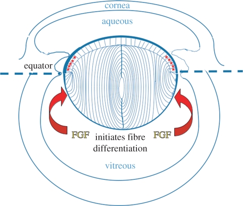
Growth factors play key roles in influencing cell fate and behaviour during development. The epithelial cells and fibre cells that arise from the lens vesicle during lens morphogenesis are bathed by aqueous and vitreous, respectively. Vitreous has been shown to generate a high level of fibroblast growth factor (FGF) signalling that is required for secondary lens fibre differentiation. However, studies also show that FGF signalling is not sufficient and roles have been identified for transforming growth factor-β and Wnt/Frizzled families in regulating aspects of fibre differentiation. In the case of the epithelium, key roles for Wnt/β-catenin and Notch signalling have been demonstrated in embryonic development, but it is not known if other factors are required for its formation and maintenance. This review provides an overview of current knowledge about growth factor regulation of differentiation and maintenance of lens cells. It also highlights areas that warrant future study.


Figures

Figure 1.
Figure 1.
Diagram of the rodent eye showing the structure of the fully formed lens. Epithelial cells and fibre cells are contained within a capsule of extracellular matrix (thick blue line). The germinative zone (red asterisks) is situated just above the equator. Cells that shift below the equator are exposed to the relatively high level of fibroblast growth factor (FGF) in vitreous and initiate a signalling cascade that promotes fibre differentiation.
Figure 2.
Figure 2.
Growth factor-induced signalling profiles of ERK1/2 and pAkt phosphorylation in lens epithelial cells. (a). Representative western blots of rat lens epithelial explants treated with either: (i) 50% bovine aqueous; (ii) 5 ng ml−1 FGF-2; (iii) 50% bovine vitreous; or (iv) 100 ng ml−1 FGF-2, and assayed for phosphorylated ERK1/2 (upper panels) or total ERK1/2 (lower panels) over a 24 h period. The first lanes indicate control explants assayed at 24 h (c24 h). (b) Summary of the duration of ERK1/2 (dark grey bars) and Akt (light grey bars) phosphorylation over a 24 h period, in cultured explants of rat lens epithelial cells exposed to different treatments. Each treatment is scored for its ability to induce secondary lens fibre differentiation in vitro: –, no response; +, weak response with little multi-layering and some β-crystallin accumulation; ++, intermediate response with notable multi-layering, some cell elongation and β-crystallin accumulation; +++, strong response with extensive multi-layering, cell elongation and abundant β-crystallin accumulation (hash denotes the study of [26]; asterisk denotes 100 ng ml−1; dagger denotes 5 ng ml−1). Adapted from Wang et al. [27].
Figure 3.
Figure 3.
Role of activin and TGF-β receptors in fibre differentiation. Null mutations of Acvr2b (ad) or Tgfbr2 (e, f) do not affect lens development. However, expression of a dominant-negative Tgfbr2 receptor results in the failure of terminal lens fibre differentiation (h). (ad); Tissues courtesy of Dr S. Paul Oh, de Iongh, 2004, unpublished data, University of Florida, Gainesville, FL, USA. (e,f) Reproduced and adapted with permission from Beebe et al. [55]. (g,h) Adapted from de Iongh et al. [37].
Figure 4.
Figure 4.
Wnt/β-catenin signalling regulates epithelial cell phenotype, proliferation and differentiation in embryonic lenses. Tissues were stained with either haematoxylin and eosin (a,d,g), E-cadherin antibody (b,e,h) or for BrdU-incorporation (c,f,i). (ac) At E13.5, cell proliferation in wild-type lens (Wt) is restricted to the E-cadherin-positive epithelial cells (c, arrowheads). (df) Loss of β-catenin (CatnbEx3-6/Ex3-6/Cre) abrogates Wnt signalling and results in the loss of lens epithelial cells (e, arrowheads) and reduction in lens cell proliferation (f, arrowheads). (gi) Constitutive activation of Wnt/β-catenin signalling by a truncating mutation of Apc (Apc580S/580S/Cre) results in the failure of lens equatorial cells to differentiate into fibres, with aberrant labelling for E-cadherin (h) and cell proliferation (i) extending into the fibre cell mass. Adapted from Cain et al. [43] and Martinez et al. [44].
Figure 5.
Figure 5.
Centrosome/cilium polarization in lens cells. (a) Transmission electron microscopy of the boxed area in the diagram (top right inset) shows the location of three adjoining cortical fibres in the lens cortex that display basal bodies (arrows) at their apical ends. The basal body of one fibre cell includes a section through its ciliary axoneme (middle arrow). A transverse section through basal bodies shows that central microtubules are absent (top left inset) and identifies them as primary cilia. (b) Pericentrin (green) immuno-reactivity localizes to the centrosome/cilium, and β-catenin (purple) localization demarcates cell margins in lens whole mounts. The centrosome/cilium is clearly associated with the cell margin proximal to the anterior pole. Scale bar; (a) 2 µm; (a, top left inset) 0.7 µm; (b), 20 µm; (b, inset) 10 µm. Adapted from Sugiyama et al. [84].
Figure 6.
Figure 6.
Histological analysis of embryonic and neonatal Sfrp2 transgenic lenses. Lens sections from E14.5 (a,b), E16.5 (c,d) and P1 (e,f) Sfrp2-m2 transgenic mice (b,d,f) and wild-type (WT) littermates (a,c,e) stained with haematoxylin and eosin. (a,b) At E14.5, compared with the WT lens, the lenses in Sfrp2-m2 mice showed essentially normal features in the epithelial and fibre cell compartments. (c,d) At E16.5 the Sfrp2-m2 lenses were slightly smaller than lenses from WT littermates. The secondary fibres in the Sfrp2-m2 lenses appeared to be less closely packed. Moreover, fibres did not curve towards the developing sutures as they did in WT lenses (black lines indicate the curvature of fibre cells in all figures). In WT lenses, the elongating fibres had a concave curvature in the transitional zone but as they moved centrally they progressively developed a convex curvature (arrowheads in c). When fibres met up with equivalent fibres from another segment of the lens, they formed rudimentary sutures (asterisks in c). In the Sfrp2-m2 lenses, the fibres retained a concave curvature and no sutures were evident. (e,f) At P1 in the transitional zone just below the lens equator (small arrow in e) the elongating fibres had a concave curvature but took on a convex curvature as they migrated at their anterior and posterior tips (large arrows) towards the anterior and posterior poles of the lens, respectively. In the sfrp2-m2 lenses, the concave curvature of the fibres was more pronounced than at earlier stages. The elongating fibres in the Sfrp2-m2 lenses remained predominantly at right angles to the posterior capsule and to the epithelium (arrowheads in f). The insets in (e) and (f) show more details of the alignment of the fibres (arrows) and the capsule (red) in WT and Sfrp2-m2 lenses. No sutures formed in the Sfrp2-m2 lenses. tz, Transitional zone. Adapted from Chen et al. [42].
Figure 7.
Figure 7.
Lrp6−/− mutant mouse: sagittal sections of an eye from an Lrp6−/− mutant embryo (a) and a wild-type embryo (b) at E14.5. Histological sections stained with haematoxylin and phloxine show that the embryonic eye in the Lrp6−/− mutant is smaller than the wild-type. The lens epithelium is incompletely formed and elongating lens fibres have extruded into the overlying cornea (asterisks). The inset in (a) confirms that the extruded material contains fibre-specific β-crystallin. Adapted from Stump et al. [109].

References

    1. McAvoy J. W. 1978. Cell division, cell elongation and distribution of alpha-, beta- and gamma-crystallins in the rat lens. J. Embryol. Exp. Morphol. 44, 149–165 - PubMed
    1. Coulombre J. L., Coulombre A. J. 1963. Lens development: fiber elongation and lens orientation. Science 142, 1489–1490 10.1126/science.142.3598.1489 (doi:10.1126/science.142.3598.1489) - DOI - PubMed
    1. Yamamoto Y. 1976. Growth of lens and ocular environment: role of neural retina in the growth of mouse lens as revealed by an implantation experiment. Dev. Growth Differ. 18, 273–278 10.1111/j.1440-169X.1976.00273.x (doi:10.1111/j.1440-169X.1976.00273.x) - DOI - PubMed
    1. Lovicu F. J., McAvoy J. W. 2005. Growth factor regulation of lens development. Dev. Biol. 280, 1–14 10.1016/j.ydbio.2005.01.020 (doi:10.1016/j.ydbio.2005.01.020) - DOI - PubMed
    1. West-Mays J. A., Pino G., Lovicu F. J. 2009. Development and use of the lens epithelial explant system to study lens differentiation and cataractogenesis. Prog. Retin. Eye Res. 29, 135–143 10.1016/j.preteyeres.2009.12.001 (doi:10.1016/j.preteyeres.2009.12.001) - DOI - PMC - PubMed

Publication types

MeSH terms