Review
. 2011 Apr 27;366(1568):1278-92.
doi: 10.1098/rstb.2010.0300.

The ageing lens and cataract: a model of normal and pathological ageing

Affiliations
Review

The ageing lens and cataract: a model of normal and pathological ageing

R Michael et al. Philos Trans R Soc Lond B Biol Sci. .

Abstract

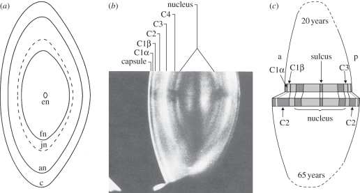
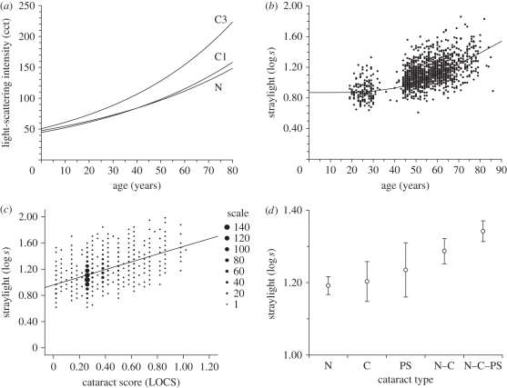
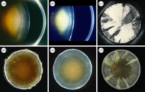
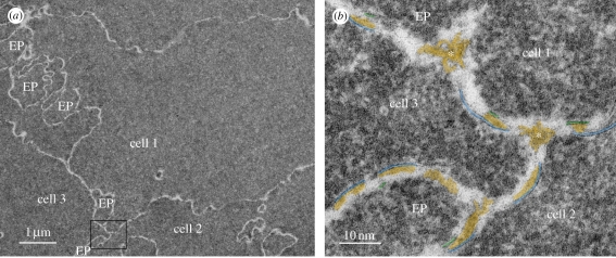
Cataract is a visible opacity in the lens substance, which, when located on the visual axis, leads to visual loss. Age-related cataract is a cause of blindness on a global scale involving genetic and environmental influences. With ageing, lens proteins undergo non-enzymatic, post-translational modification and the accumulation of fluorescent chromophores, increasing susceptibility to oxidation and cross-linking and increased light-scatter. Because the human lens grows throughout life, the lens core is exposed for a longer period to such influences and the risk of oxidative damage increases in the fourth decade when a barrier to the transport of glutathione forms around the lens nucleus. Consequently, as the lens ages, its transparency falls and the nucleus becomes more rigid, resisting the change in shape necessary for accommodation. This is the basis of presbyopia. In some individuals, the steady accumulation of chromophores and complex, insoluble crystallin aggregates in the lens nucleus leads to the formation of a brown nuclear cataract. The process is homogeneous and the affected lens fibres retain their gross morphology. Cortical opacities are due to changes in membrane permeability and enzyme function and shear-stress damage to lens fibres with continued accommodative effort. Unlike nuclear cataract, progression is intermittent, stepwise and non-uniform.


Figures

Figure 1.
Figure 1.
(a) Diagram of the lens zones drawn to scale based on a lens equatorial diameter of 9.6 mm in meridional section. The embryonic nucleus (en) represents the size of the fibre mass at the end of embryonic life; the foetal nucleus (fn) is the size of the fibre mass at the time of birth. This is referred to as the ‘true nucleus’ in this paper. The terms juvenile nucleus (jn) and adult nucleus (an) are also used in the literature to convey, respectively, those parts of the lens mass formed by the end of the second decade and later in adult life [38]. However, this terminology is not used here. (b) Scheimpflug image of an adult normal lens to show the zones according to the Oxford Classification (see text for details) [37]. (c) Schematic of the zones in the lens of a 20-year-old subject (upper part) and a 65-year-old subject to indicate lens growth with ageing. As the lens grows, only C2 is seen to increase in thickness. The ‘sulcus’ of the lens refers to the drop in light-scattering that occurs at the very centre of the lens (in the region of the embryonic nucleus). The ‘true nucleus’ includes the sulcus and the light grey zone on either side. The dark band immediately outside the true nucleus corresponds to C4 of the Oxford grading system. Note that Dubbelman includes C4 in his descriptions of the nucleus [39]. Figure 1a from Taylor et al. [38], © 1996 by Investigative Ophthalmology & Visual Science. Adapted from Investigative Ophthalmology & Visual Science in the format Journal via Copyright Clearance Center. Figure 1b reprinted from Sparrow et al. [37] with kind permission from Springer Science + Business Media. Figure 1c reprinted from Dubbelman et al. [39], © 2003, with permission from Elsevier.
Figure 2.
Figure 2.
(a) Densitometric evaluation of Scheimpflug images to measure backward light-scattering from the lens and (bd) psychophysical measurements of forward intraocular light-scattering. Light-scattering is shown as a function of (a,b) age, (c) the cataract score and (d) cataract type. (a) C1, C3 and N (nucleus) are lens zones according to figure 1b. (d) N, nuclear; C, cortical; PS, posterior subcapsular; N–C, mixed nuclear and cortical; N–C–PS, mixed nuclear, cortical and posterior subcapsular. Error bars show the confidence interval for the mean. Figure 2a reprinted from Sasaki [41], with permission from Deutsche Akademie der Naturforscher Leopoldina—Nationale Akademie der Wissenschaften. Figure 2b reprinted from van den Berg et al. [42], © 2007, with permission from Elsevier. Figure 2c reprinted from Michael et al. [43], with permission from John Wiley and Sons. Figure 2d reprinted from Nischler et al. [44], with permission from Wichtig Editore Srl.
Figure 3.
Figure 3.
(a,d) Brunescent nuclear cataract of moderate-to-marked density. The deep cortex is also coloured a yellowish brown. In (a) the broad light-scattering zone in the deep cortex is C3. Note that the cortical zone, C1, is intact. (b,e) A dense, non-brunescent, white nuclear cataract. Anterior and posterior subcapsular cataracts are also present. (a,b) Scheimpflug photography (courtesy of J. M. Sparrow) with the cornea seen to the right. (d,e) Dark-field micrographs of human donor lenses. (c,f) Spoke cataract of varying density and extent, viewed by retroillumination in a living patient (c) and dark-field illumination of an extracted lens (f); in neither case is there an associated nuclear cataract. Scheimpflug and retroillumination images (ac) and dark-field micrographs (df) are from different subjects. Figure 3d reprinted from Michael et al. [64], © 2008, with permission from Elsevier.
Figure 4.
Figure 4.
Ultrastructural images of fibre, cell–cell and cell–process interactions, in the nucleus of an advanced nuclear cataract from a 51-year-old patient from India. (a) The interfaces between three fibre cells and several edge processes (EP) are shown. (b) High-magnification view of the region boxed in (a), revealing damaged membranes from three cells and two edge processes. Extracellular space deposits (yellow) appear on curved membranes (blue and green lines) and similarly staining material occurs at trigonal intersections (asterisk). The trigonal points are extended extracellular channels that are partially filled with protein-like material. Figure 4 from Costello et al. [28], © 2008 by Elsevier Science and Technology Journals. Adapted from Elsevier Science & Technology Journals in the format Journal via Copyright Clearance Center.
Figure 5.
Figure 5.
Dark-field micrographs of aged human donor lenses, illustrating (a) small, dot-like opacities and (b) radial and circular shades. (c) Multilamellar body, as frequently found in human lenses with early cortical opacities probably causing the star-like opacities seen in (a). A slice cut in the axial plane of the fixed donor lens (b) is shown in (d). Arrows, radial shades and arrow heads, circular shades. Figure 5a,b reprinted from Michael et al. [64], © 2008, with permission from Elsevier. Figure 5c reprinted from Vrensen et al. [70], with permission from Taylor & Francis. Figure 5d reprinted from Michael [71], © 2010, with permission from Elsevier. Scale bars, (a,b) 500 µm, (c) 1 µm, (d) 1000 µm.
Figure 6.
Figure 6.
(ac) Panel of dark-field micrographs of aged human donor lenses and (df) of slices cut in the axial plane of the fixed donor lenses. The cutting plane is indicated by a white line in (ac). Apart from some irregular scattering owing to imperfect slicing, the nuclear parts of the slices are free of opacification. In (b) a band-like opacity is seen. Except for the spokes in (c), the opacities are outside the pupillary space and will not have seriously influenced vision. Below is the fibre organization in a lens without cortical cataract and in two cases of cortical opacities (boxed areas in df), as visualized by scanning electron microscopy (gi). (h) Fibres at the border zone between the C3 and C2 regions are broken (arrows) and the broken ends are directed towards the nuclear fibres, which maintain regular, uninterrupted organization. Further, note the curled (asterisk) and folded (arrowheads) fibres in the region adjoining the broken fibres. (i) The scanning electron microscopy micrograph shows broken fibres at several places (arrows) in the border zone between the C3 and C2 regions. The central fibres are regularly organized, as are the more superficial cortical fibres bridging the break zone. ep, epithelium; n, nuclear side; c, cortical side. Scale bars, (df,i) 1000 µm and (g,h) 100 µm. Figure 6 reprinted from Michael et al. [64], © 2008, with permission from Elsevier.

References

    1. Winkler B. S., Riley M. V. 1991. Relative contributions of epithelial cells and fibers to rabbit lens ATP content and glycolysis. Invest. Ophthalmol. Vis. Sci. 32, 2593–2598 - PubMed
    1. Mathias R. T., White T. W., Gong X. 2010. Lens gap junctions in growth, differentiation, and homeostasis. Physiol. Rev. 90, 179–20610.1152/physrev.00034.2009 (doi:10.1152/physrev.00034.2009) - DOI - DOI - PMC - PubMed
    1. Augusteyn R. C. 1981. Protein modification in cataract: possible oxidative mechanism. In Mechanisms of cataract formation in the human lens (ed. Duncan G.), pp. 72–115 New York, NY: Academic Press
    1. Harding J. J. 1991. Cataract: biochemistry, epidemiology and pharmacology. London, UK: Chapman & Hall
    1. Lou M. F. 2003. Redox regulation in the lens. Prog. Retin. Eye Res. 22, 657–68210.1016/S1350-9462(03)00050-8 (doi:10.1016/S1350-9462(03)00050-8) - DOI - DOI - PubMed

MeSH terms