. 2011 May 13;286(19):17156-67.
doi: 10.1074/jbc.M111.231571. Epub 2011 Mar 14.

ADAMTS10 protein interacts with fibrillin-1 and promotes its deposition in extracellular matrix of cultured fibroblasts

Affiliations

ADAMTS10 protein interacts with fibrillin-1 and promotes its deposition in extracellular matrix of cultured fibroblasts

Wendy E Kutz et al. J Biol Chem. .

Abstract

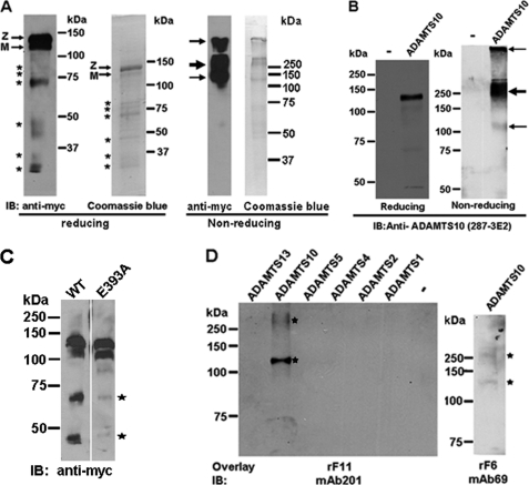
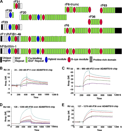
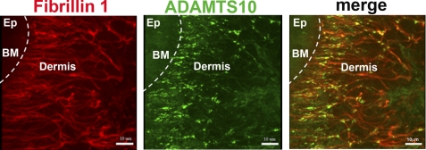
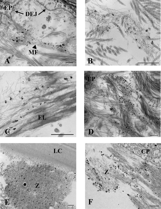
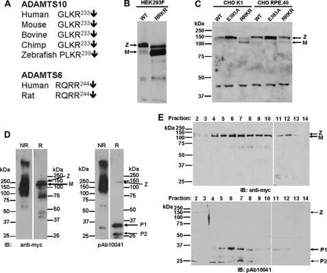
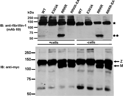
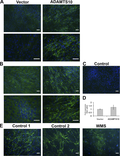
Autosomal recessive and autosomal dominant forms of Weill-Marchesani syndrome, an inherited connective tissue disorder, are caused by mutations in ADAMTS10 (encoding a secreted metalloprotease) and FBN1 (encoding fibrillin-1, which forms tissue microfibrils), respectively, yet they are clinically indistinguishable. This genetic connection prompted investigation of a potential functional relationship between ADAMTS10 and fibrillin-1. Specifically, fibrillin-1 was investigated as a potential ADAMTS10 binding partner and substrate, and the role of ADAMTS10 in influencing microfibril biogenesis was addressed. Using ligand affinity blotting and surface plasmon resonance, recombinant ADAMTS10 was found to bind to fibrillin-1 with a high degree of specificity and with high affinity. Two sites of ADAMTS10 binding to fibrillin-1 were identified, one toward the N terminus and another in the C-terminal half of fibrillin-1. Confocal microscopy and immunoelectron microscopy localized ADAMTS10 to fibrillin-1-containing microfibrils in human tissues. Furin-activated ADAMTS10 could cleave fibrillin-1, but innate resistance of ADAMTS10 zymogen to propeptide excision by furin was observed, suggesting that, unless activated, ADAMTS10 is an inefficient fibrillinase. To investigate the role of ADAMTS10 in microfibril biogenesis, fetal bovine nuchal ligament cells were cultured in the presence or absence of ADAMTS10. Exogenously added ADAMTS10 led to accelerated fibrillin-1 microfibril biogenesis. Conversely, fibroblasts obtained from a Weill-Marchesani syndrome patient with ADAMTS10 mutations deposited fibrillin-1 microfibrils sparsely compared with unaffected control cells. Taken together, these findings suggest that ADAMTS10 participates in microfibril biogenesis rather than in fibrillin-1 turnover.


Figures

FIGURE 1.
FIGURE 1.
ADAMTS10 binds to fibrillin-1. A, analysis of purified recombinant ADAMTS10 by Western blot analysis (under reducing and non-reducing conditions as indicated) with anti-myc antibody or Coomassie Blue staining. Under reducing conditions, the major ADAMTS10 species migrates at 130 kDa, consistent with it being the unprocessed zymogen (Z). A 120-kDa band corresponding to mature (M) ADAMTS10 is present. Additional, smaller immunoreactive bands seen (asterisks) appear to be derived from ADAMTS10 on the basis of their binding to anti-myc. Under non-reducing conditions, the major species detected is at 250 kDa (bold arrow) with minor species seen at 120 kDa and within the stacking gel (fine arrows). B, characterization of monoclonal antibody 287-3E2 by Western blot analysis of conditioned medium from ADAMTS10-transfected HEK293F cells. Transfection with the pcDNA vector alone was used as the control (−). Under reducing conditions, the expected 130-kDa zymogen (Z; a major species) and the 120-kDa mature form (M; a minor species) are specifically seen only in the medium of ADAMTS10-transfected cells. Under non-reducing conditions, most of the ADAMTS10 migrates with an apparent molecular mass of 250 kDa (bold arrow). Some is trapped in the stacking gel, and a small amount with an apparent molecular mass of 120 kDa is also seen (fine arrows). C, wild type (WT) or ADAMTS10 E393A was transiently transfected in parallel into HEK293F cells, and the collected medium was electrophoresed under reducing conditions followed by immunoblotting with anti-myc. Note that molecular species corresponding to proteolytic fragments in the WT sample (asterisks) are considerably weaker in the E393A medium. D, left-hand panel, 400 ng each of ADAMTS10 and the indicated ADAMTS proteases were electrophoresed under non-reducing conditions. Following electroblotting, membranes were incubated with fibrillin-1 peptide corresponding to the N-terminal half (rF11) followed by detection with the indicated fibrillin-1 antibody. The peptide bound only to ADAMTS10 (asterisks). Right-hand panel, ADAMTS10 was electrophoresed as above for investigation of interaction with the C-terminal half of fibrillin-1 (rF6) followed by detection using mAb 69. The ADAMTS10 species detected were similar to those that bound rF11 (asterisks). IB, immunoblot.
FIGURE 2.
FIGURE 2.
SPR analysis of interactions of full-length ADAMTS10 with fibrillin-1. A, depiction of the domain structure of fibrillin-1 and the peptides used in SPR analysis. B–E, the indicated fibrillin-1 peptides were used as the analyte and flowed over a chip coupled with ADAMTS10 as the ligand. The observed SPR change is indicated in resonance units (RU). Injections utilized a series of molar concentrations for each analyte (the concentration range used is indicated above each set of sensorgrams). Table 1 lists the specific numerical values for binding obtained from these and additional experiments of which the illustrated data are representative. Resp. Diff., response difference.
FIGURE 3.
FIGURE 3.
Co-localization of ADAMTS10 with fibrillin-1 in human skin. A, immunofluorescent visualization of the distribution of fibrillin-1 (red) and ADAMTS10 (green) in human skin. The locations of the epidermis (Ep), dermis, and basement membrane (BM; broken white line) are shown. Note the substantial overlap between ADAMTS10 and fibrillin microfibrils in the papillary dermis (closest to the basement membrane) with relatively lower ADAMTS10 staining intensity in the further removed reticular dermis (center and right of each panel). Scale bar, 10 μm. Negative controls indicating antibody specificity are shown in the supplemental data.
FIGURE 4.
FIGURE 4.
Immunoelectron microscopy illustrates that ADAMTS10 is specifically bound to tissue microfibrils in skin and zonules. ADAMTS10 was localized en bloc using mAb 287-3E2 followed by 1-nm gold secondary conjugate and then gold-enhanced to allow visualization at low magnification. Labeling of microfibril bundles (MF) is most intense close to the dermal-epidermal junction; however, labeling is absent immediately adjacent to the epithelium (EP) and the basement membrane at the dermal-epidermal junction (DEJ; A, arrows). Immunogold particles seen at higher magnification following slightly less time in gold enhancement solution demonstrate that ADAMTS10 localizes to microfibrils in small clumps represented by a cluster of gold particles (B; image taken near the dermal-epidermal junction) also seen among elastin-associated microfibrils in the shallow reticular dermis (C). An image collected at 0° tilt through an exceptionally thick section (300 nm; D) of immunolabeled skin shows several labeled microfibril bundles intersecting the lamina densa of the dermal-epidermal junction best appreciated in the aligned tilt series (supplemental Video File 2). Ciliary zonules (Z) intersecting both the lens capsule (LC; E) and ciliary process (CP; F) are well labeled with antibody to ADAMTS10. Scale bars for A–D, 500 nm; scale bars for E and F, 1 μm.
FIGURE 5.
FIGURE 5.
Wild-type ADAMTS10 is normally resistant to furin processing but can be substantially processed to mature form by optimization of furin recognition sequence. A, sequence alignment of the putative furin cleavage site of ADAMTS10 and its closest homolog, ADAMTS6, from the indicated species. Note that the cleavage site of ADAMTS6 matches the desired RX(K/R)R consensus but that ADAMTS10 consistently lacks the P4 Arg residue. B, site-directed mutagenesis of ADAMTS10 was used to convert the wild-type (WT) G230L231KR sequence to R230R231KR (RRKR), and both forms were analyzed by Western blot of the conditioned medium of transfected HEK293F cells. Note that the major species detected in WT ADAMTS10 is the zymogen (Z), whereas the mature form (M) constitutes the major species following ADAMTS10-RRKR transfection. C, analysis of the indicated ADAMTS10 constructs in medium of transiently transfected CHO-K1 cells or furin-deficient CHO-RPE.40 cells by reducing SDS-PAGE and Western blotting with anti-myc. Note that the prevalent mature form (M) of the ADAMTS10-RRKR mutant is decreased in intensity relative to the zymogen (Z) in CHO-RPE.40, cells indicating that it was the consequence of furin processing. In contrast, there is little change in amount of the mature and zymogen forms observed in wild-type ADAMTS10 (WT) or ADAMTS10 E393A in either cell line. D, comparison of ADAMTS10-RRKR migration under reducing (R) and non-reducing (NR) conditions using anti-myc or the anti-propeptide antibody pAb 10041. Note the similar migration pattern observed with both antibodies under non-reducing conditions with the exception of some cleaved propeptide observed with pAb 10041. In contrast, reducing Western blots with anti-myc show a prevalent 120-kDa band corresponding to mature ADAMTS10 (M) with a low prevalence of zymogen (Z), and pAb 10041 showed prominent species of 35 (P1) and 27 kDa (P2) arising from the processed ADAMTS10 propeptide. E, Western blots following reducing SDS-PAGE of fractions collected from analytical gel filtration chromatography of ADAMTS10-RRKR. Note that cleaved ADAMTS10 propeptide species P1 and P2 co-elute with the mature ADAMTS10 in fractions 4–12. IB, immunoblot.
FIGURE 6.
FIGURE 6.
Furin-processed ADAMTS10 can cleave fibrillin-1. ADAMTS10 proteolytic activity toward fibrillin-1 was determined in HEK293F cells stably transfected with wild-type ADAMTS10 (WT) or the indicated mutants. The transfected cells (+cells) or their conditioned medium (−cells) as indicated was incubated with peptide rF6 (asterisk). ADAMTS10 (WT) cleaves rF6 to generate a 60-kDa peptide (top panel, double asterisk), but this cleavage is much more robust with ADAMTS10-RRKR. When rF6 digests were done using ADAMTS10-conditioned cell-free medium, cleavage by wild-type ADAMTS10 was undetectable. Mutation of the ADAMTS10 catalytic Glu393 residue (to Ala) in each construct abrogated cleavage and is indicative of specific cleavage by ADAMTS10 in this experiment. The lower panel shows the ADAMTS10 molecular species detected by Western blotting with anti-myc. IB, immunoblot; Z, zymogen; M, mature form.
FIGURE 7.
FIGURE 7.
ADAMTS10 accelerates microfibril biogenesis in cultured fibroblasts, and WMS fibroblasts assemble a sparse microfibril network compared with normal skin fibroblasts. A and B, panels show fibrillin-1 immunofluorescence (green) in cultures of bovine fetal nuchal ligament cells as described under “Experimental Procedures.” Two fields are shown from cultures grown for 3 (A) or 5 days (B) at two different magnifications and are representative of multiple (n = 10) independent experiments. The cells treated with ADAMTS10-conditioned medium consistently show more fibrillin-1 microfibrils than cells treated with vector-transfected conditioned medium. In the negative control panel (C), the anti-fibrillin-1 antibody was omitted, and this preparation showed no green fluorescence. D, quantitative RT-PCR of FBN1 mRNA from fBNL fibroblasts treated with ADAMTS10-conditioned medium or vector-conditioned medium (n = 3) shows that there was no significant difference. Error bars represent S.D. E, skin fibroblasts from two control male subjects and an 86-year-old man with WMS were grown to confluence and stained for fibrillin-1 microfibrils (green) using monoclonal antibody 69. Note the robust microfibril production in cells from the control subjects, whereas fewer microfibrils were present in the WMS fibroblasts after 5 days. Scale bar, 50 μm.

Similar articles

Cited by

References

    1. Sakai L. Y., Keene D. R., Engvall E. (1986) J. Cell Biol. 103, 2499–2509 - PMC - PubMed
    1. Ramirez F., Sakai L. Y., Dietz H. C., Rifkin D. B. (2004) Physiol. Genomics 19, 151–154 - PubMed
    1. Cain S. A., Morgan A., Sherratt M. J., Ball S. G., Shuttleworth C. A., Kielty C. M. (2006) Proteomics 6, 111–122 - PubMed
    1. Pyeritz R. E. (2000) Annu. Rev. Med. 51, 481–510 - PubMed
    1. Judge D. P., Biery N. J., Keene D. R., Geubtner J., Myers L., Huso D. L., Sakai L. Y., Dietz H. C. (2004) J. Clin. Investig. 114, 172–181 - PMC - PubMed

Publication types