. 2011 Apr 11;208(4):643-51.
doi: 10.1084/jem.20100682. Epub 2011 Mar 14.

Nucleic acid recognition by Toll-like receptors is coupled to stepwise processing by cathepsins and asparagine endopeptidase

Affiliations

Nucleic acid recognition by Toll-like receptors is coupled to stepwise processing by cathepsins and asparagine endopeptidase

Sarah E Ewald et al. J Exp Med. .

Abstract

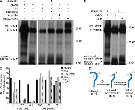
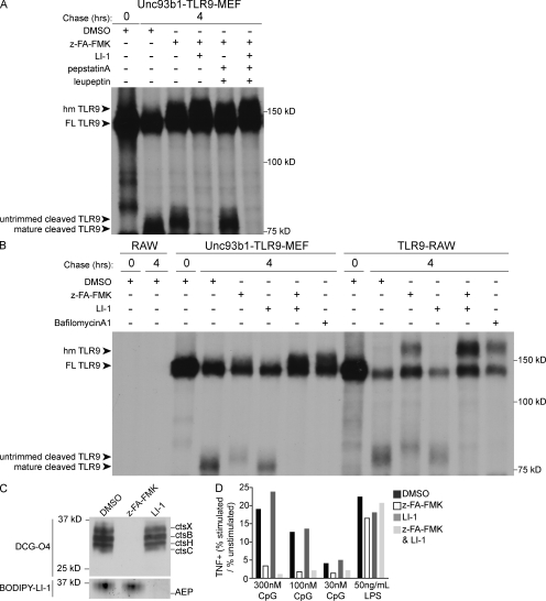
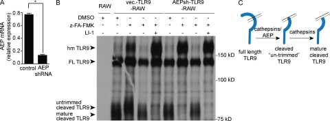
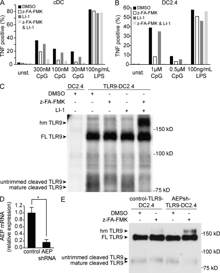
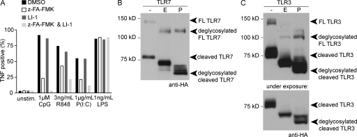
Toll-like receptor (TLR) 9 requires proteolytic processing in the endolysosome to initiate signaling in response to DNA. However, recent studies conflict as to which proteases are required for receptor cleavage. We show that TLR9 proteolysis is a multistep process. The first step removes the majority of the ectodomain and can be performed by asparagine endopeptidase (AEP) or cathepsin family members. This initial cleavage event is followed by a trimming event that is solely cathepsin mediated and required for optimal receptor signaling. This dual requirement for AEP and cathepsins is observed in all cell types that we have analyzed, including mouse macrophages and dendritic cells. In addition, we show that TLR7 and TLR3 are processed in an analogous manner. These results define the core proteolytic steps required for TLR9 function and suggest that receptor proteolysis may represent a general regulatory strategy for all TLRs involved in nucleic acid recognition.


Figures

Figure 1.
Figure 1.
TLR9 cleavage is a multistep event. (A and B) TLR9 cleavage in TLR9-RAW cells was monitored by pulse-chase analysis in the presence of the indicated protease inhibitors or DMSO (as vehicle control). The full-length form of TLR9 (FL TLR9) and two cleavage products (mature cleaved TLR9 and untrimmed cleaved TLR9) are labeled. The pool of full-length TLR9 that has exited the ER but has not yet been cleaved is also labeled (hmTLR9). (C) Analysis of TNF production by intracellular cytokine staining. RAW cells were pretreated for 12 h with the indicated protease inhibitors (broad spectrum cathepsin inhibitors [z-FA-FMK or E64d], AEP inhibitor [LI-1], a combination of ctsS inhibitor [ctsS I.], ctsL inhibitor [ctsL I.], and ctsB inhibitor [ctsB I.], or vehicle control [DMSO]) followed by 4 h of stimulation with the indicated concentrations of CpG or LPS. Graph represents the ratio of the percentage of TNF-expressing cells in stimulated and unstimulated conditions. Representative FACS plots are shown in Fig. S2. (D) Schematic depicting the unique requirement of cathepsins for trimming TLR9 once the majority of the ectodomain has been removed, generating the mature form of the cleaved receptor. All data are representative of at least five experiments.
Figure 2.
Figure 2.
AEP and cathepsins account for cleavage of the TLR9 ectodomain. (A and B) TLR9 cleavage in Unc93b1-TLR9-MEF cells or TLR9-RAW cells was monitored by pulse-chase analysis in the presence of indicated protease inhibitors or DMSO (as vehicle control). (C) Efficiency of protease inhibition of live cells was determined by probing cell lysates with tagged protease inhibitor probes. RAW cells were pretreated with z-FA-FMK (z-FA), LI-1, or DMSO. Cell lysates were then probed with the cathepsin probe DCG-04-biotin (top) or the AEP probe BODIPY-LI-1 (bottom) and visualized as described in Materials and methods. Bands corresponding to individual proteases are indicated on the right. (D) Analysis of TNF production by RAW cells using intracellular cytokine staining as described in Fig. 1. Representative FACS plots are shown in Fig. S3. All data are representative of at least three experiments.
Figure 3.
Figure 3.
In the absence of AEP, TLR9 cleavage in macrophages is entirely cathepsin dependent. (A) Quantitative RT-PCR analysis of AEP transcript levels in TLR9-RAW cells transduced with an shRNA construct targeting AEP (AEP-shRNA) or vector control. AEP levels were normalized to rps17 expression. Error bars represent standard deviation. * represents P ≤ 0.001 based on Student’s t test. (B) TLR9 cleavage in the cells described in A as analyzed by pulse-chase analysis in the presence of indicated protease inhibitors (z-FA-FMK; LI-1) or DMSO (as vehicle control). All data are representative of two experiments. (C) Schematic illustrating the overlapping roles of AEP and cathepsins in the initial removal of the TLR9 ectodomain.
Figure 4.
Figure 4.
TLR9 signaling and cleavage in DCs requires AEP and cathepsins. (A and B) TNF production by GM-CSF–derived DCs (A) or DC2.4 cells (B) was monitored by intracellular cytokine staining as described in Fig. 1. Representative FACS plots are shown in Fig. S4. Data are representative of three experiments. (C) TLR9 cleavage in TLR9-DC2.4 cells or DC2.4 cells was monitored by pulse-chase analysis as described in Materials and methods. Data are representative of four experiments. (D) Quantitative RT-PCR analysis of AEP transcript levels in TLR9-DC2.4 cells transduced with the AEP shRNA (AEP-shRNA) or an unrelated gene product (control). AEP levels were normalized to rps17 expression. Error bars represent standard deviation. * represents P ≤ 0.005 based on Student’s t test. (E) TLR9 cleavage in the cells described in D visualized by immunoblot after immunoprecipitation after 14 h of z-FA-FMK or DMSO treatment. Data in E are representative of three experiments.
Figure 5.
Figure 5.
TLR7 and TLR3 are regulated by receptor proteolysis. (A) TNF production by RAW cells in response to TLR9, TLR3, and TLR7 ligands was measured by intracellular cytokine staining as described in Fig. 1. Representative FACS plots are shown in Fig. S6. (B and C) TLR7 (B) and TLR3 (C) are cleaved after trafficking through the Golgi. TLR7-RAW (B) or TLR3-RAW (C) cell immunoprecipitates were treated with Endo H (E), PNGase (P), or no enzyme control (−) to assess glycosylation status by anti-HA Western blotting. The bottom panel in C is a shorter exposure of the TLR3 cleaved product. All data are representative of three experiments.

References

    1. Asagiri M., Hirai T., Kunigami T., Kamano S., Gober H.-J., Okamoto K., Nishikawa K., Latz E., Golenbock D.T., Aoki K., et al. 2008. Cathepsin K-dependent toll-like receptor 9 signaling revealed in experimental arthritis. Science. 319:624–627 10.1126/science.1150110 - DOI - PubMed
    1. Barton G.M., Kagan J.C., Medzhitov R. 2006. Intracellular localization of Toll-like receptor 9 prevents recognition of self DNA but facilitates access to viral DNA. Nat. Immunol. 7:49–56 10.1038/ni1280 - DOI - PubMed
    1. Bell J.K., Askins J., Hall P.R., Davies D.R., Segal D.M. 2006. The dsRNA binding site of human Toll-like receptor 3. Proc. Natl. Acad. Sci. USA. 103:8792–8797 10.1073/pnas.0603245103 - DOI - PMC - PubMed
    1. Botos I., Liu L., Wang Y., Segal D.M., Davies D.R. 2009. The toll-like receptor 3:dsRNA signaling complex. Biochim. Biophys. Acta. 1789:667–674 - PMC - PubMed
    1. Brinkmann M.M., Spooner E., Hoebe K., Beutler B., Ploegh H.L., Kim Y.-M. 2007. The interaction between the ER membrane protein UNC93B and TLR3, 7, and 9 is crucial for TLR signaling. J. Cell Biol. 177:265–275 10.1083/jcb.200612056 - DOI - PMC - PubMed

Publication types