. 2011 Apr 11;208(4):633-41.
doi: 10.1084/jem.20110251. Epub 2011 Mar 14.

Mechanisms of necroptosis in T cells

Affiliations

Mechanisms of necroptosis in T cells

Irene L Ch'en et al. J Exp Med. .

Abstract

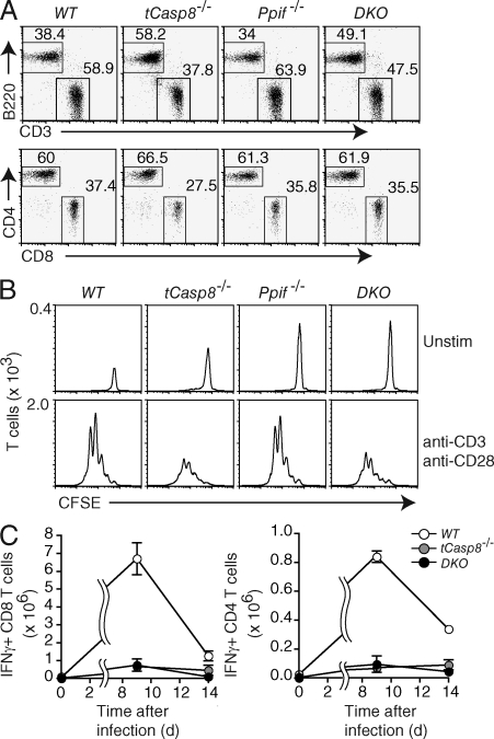
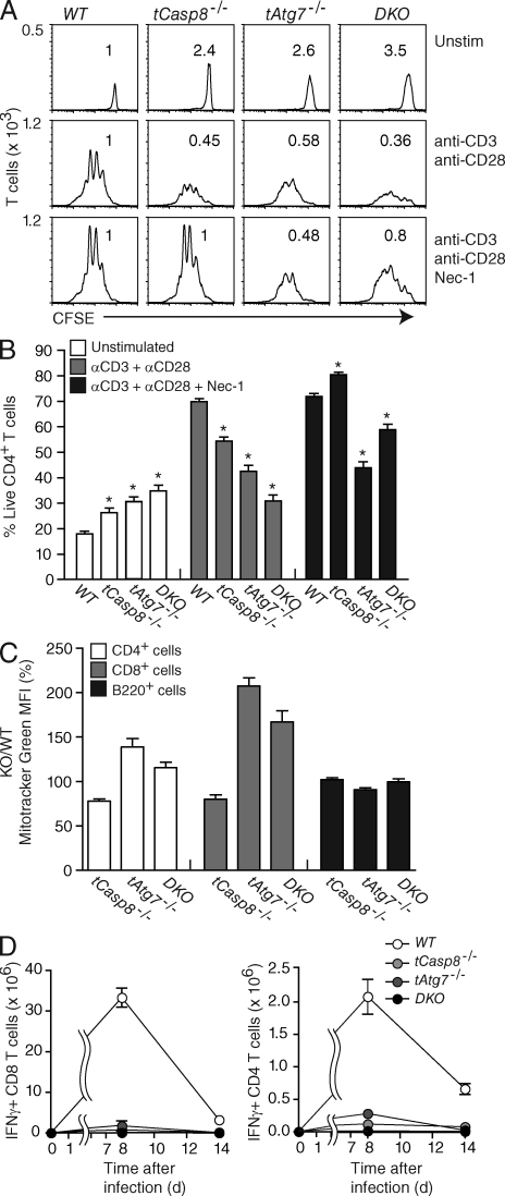
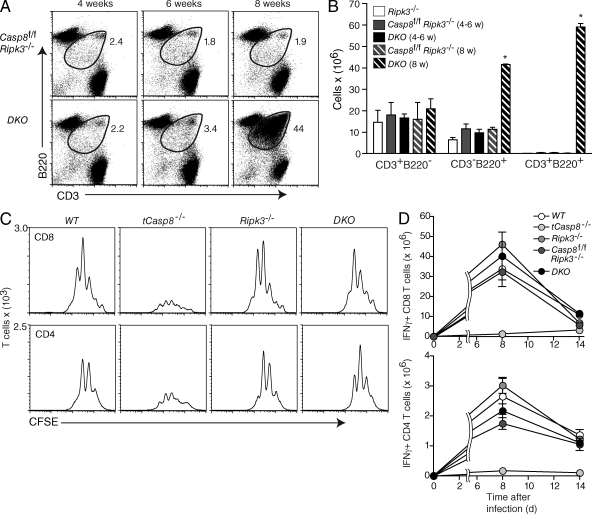
Cell populations are regulated in size by at least two forms of apoptosis. More recently, necroptosis, a parallel, nonapoptotic pathway of cell death, has been described, and this pathway is invoked in the absence of caspase 8. In caspase 8-deficient T cells, necroptosis occurs as the result of antigen receptor-mediated activation. Here, through a genetic analysis, we show that necroptosis in caspase 8-deficient T cells is related neither to the programmed necrosis as defined by the requirement for mitochondrial cyclophilin D nor to autophagy as defined by the requirement for autophagy-related protein 7. Rather, survival of caspase 8-defective T cells can be completely rescued by loss of receptor-interacting serine-threonine kinase (Ripk) 3. Additionally, complementation of a T cell-specific caspase 8 deficiency with a loss of Ripk3 gives rise to lymphoproliferative disease reminiscent of lpr or gld mice. In conjunction with previous work, we conclude that necroptosis in antigen-stimulated caspase 8-deficient T cells is the result of a novel Ripk1- and Ripk3-mediated pathway of cell death.


Figures

Figure 1.
Figure 1.
Caspase 8–deficient T cells do not die by classical necrosis. (A) The percentages of live-gated T and B cells from the lymph nodes of WT (Casp8f/f), tCasp8−/− (Casp8f/f Cd4Cre), Ppif−/− (Casp8f/f Ppif−/−), and DKO (Casp8f/f Ppif−/− Cd4Cre) mice were determined by flow cytometry. Data are representative of seven independent experiments. (B) Purified T cells were labeled with CFSE, and then cultured in media alone or stimulated with anti-CD3 and anti-CD28 for 72 h. All cells were resuspended in an equal volume and collected for the same amount of time on the flow cytometer. The numbers on the ordinate indicate the number of T cells per interval of intensity, where the area under the curve equals the total number of T cells collected. Data are representative of seven independent experiments. (C) Cohorts of mice were infected with LCMV Armstrong. On days 9 and 14 after infection, mice were sacrificed, and splenocytes were stimulated with LCMV peptides for 5 h in vitro. Intracellular IFN-γ in gated CD4+ and CD8+ T cells was measured by flow cytometry. Error bars represent the SEM. Data are representative of two independent experiments.
Figure 2.
Figure 2.
Loss of Atg7 does not rescue caspase 8–deficient T cell proliferation. (A) Purified WT (Casp8f/f), tCasp8−/− (Casp8f/f Cd4Cre), tAtg7−/− (Atg7f/f Cd4Cre), and DKO (Casp8f/f Atg7f/f Cd4Cre) T cells were labeled with CFSE, and then cultured in media alone or stimulated with anti-CD3 and anti-CD28 in the absence or presence of necrostatin-1 (Nec-1) for 72 h. Flow cytometry analysis was performed as described in the legend to Fig. 1 B. Numbers above each curve represent the proportion of recovered cells relative to WT. Data are representative of eight independent experiments. (B) Percentage of live T cells was determined from proliferation described in A. Data are cumulative from eight independent experiments, and error bars indicate the SEM. Asterisks indicate a significant difference from the WT control; P < 0.01. (C) Mitochondrial mass was measured with MitoTracker Green and percent positive in tCasp8−/−, tAtg7−/−, and DKO T cells versus WT T cells was calculated. Data are cumulative from three independent experiments, and the bars indicate SEM. (D) Cohorts of mice were infected with LCMV Armstrong. On days 8 and 14 after infection, mice were sacrificed and splenocytes were stimulated with LCMV peptides for 5 h in vitro. Intracellular IFN-γ in gated CD4+ and CD8+ T cells was measured by flow cytometry. Data are representative of three independent experiments.
Figure 3.
Figure 3.
Increased apoptotic death in the absence of Atg7. Purified T cells were cultured in media alone (Unstim) or stimulated with anti-CD3 and anti-CD28 in the absence or presence of necrostatin-1 (Nec-1), qVD, or both for 72 h. Dead cells were detected by staining for Annexin V and 7AAD (A) or TUNEL and active caspase 3 (B). Genotypes are abbreviated as listed in legend to Figure 2. Data are cumulative from eight (A) or five (B) independent experiments, bars indicate the SEM, and asterisks indicate a significant difference from the WT control; P < 0.01.
Figure 4.
Figure 4.
Targeted deletion of Ripk3 rescues caspase 8–dependent necroptosis. (A) Live-gated Casp8f/f Ripk3−/− and DKO (Casp8f/f Ripk3−/− Cd4Cre) lymphocytes from mice of the indicated ages were stained with CD3 and B220. Data are representative of three independent experiments. (B) The number of live-gated CD3+B220, CD3B220+, and CD3+B220+ cells from the lymph nodes of Ripk3−/−, Casp8f/f Ripk3−/−, and DKO mice were determined by flow cytometry. Data are cumulative from three independent experiments. Bars indicate the SEM, and asterisks indicate a significant difference from the WT control; P < 0.01. (C) Purified T cells were labeled with CFSE and stimulated with anti-CD3 and anti-CD28 for 72 h. Flow cytometry analysis was performed as described in the legend to Fig. 1 B. Data are representative of three independent experiments. (D) Cohorts of mice were infected with LCMV Armstrong. On days 8 and 14 after infection, mice were sacrificed and splenocytes were stimulated with LCMV peptides for 5 h in vitro. Intracellular IFN-γ in gated CD4+ and CD8+ T cells was measured by flow cytometry. Data for days 8 and 14 after infection are representative of three and one independent experiments, respectively.

Comment in

References

    1. Arechiga A.F., Bell B.D., Solomon J.C., Chu I.H., Dubois C.L., Hall B.E., George T.C., Coder D.M., Walsh C.M. 2005. Cutting edge: FADD is not required for antigen receptor-mediated NF-kappaB activation. J. Immunol. 175:7800–7804 - PubMed
    1. Arechiga A.F., Bell B.D., Leverrier S., Weist B.M., Porter M., Wu Z., Kanno Y., Ramos S.J., Ong S.T., Siegel R., Walsh C.M. 2007. A Fas-associated death domain protein/caspase-8-signaling axis promotes S-phase entry and maintains S6 kinase activity in T cells responding to IL-2. J. Immunol. 179:5291–5300 - PubMed
    1. Baines C.P., Kaiser R.A., Purcell N.H., Blair N.S., Osinska H., Hambleton M.A., Brunskill E.W., Sayen M.R., Gottlieb R.A., Dorn G.W., et al. 2005. Loss of cyclophilin D reveals a critical role for mitochondrial permeability transition in cell death. Nature. 434:658–662 10.1038/nature03434 - DOI - PubMed
    1. Baines C.P., Kaiser R.A., Sheiko T., Craigen W.J., Molkentin J.D. 2007. Voltage-dependent anion channels are dispensable for mitochondrial-dependent cell death. Nat. Cell Biol. 9:550–555 10.1038/ncb1575 - DOI - PMC - PubMed
    1. Beisner D.R., Ch’en I.L., Kolla R.V., Hoffmann A., Hedrick S.M. 2005. Cutting edge: innate immunity conferred by B cells is regulated by caspase-8. J. Immunol. 175:3469–3473 - PubMed

Publication types

Substances