. 2011 May;79(5):1889-97.
doi: 10.1128/IAI.01257-10. Epub 2011 Mar 14.

Essential role for neutrophils in pathogenesis and adaptive immunity in Chlamydia caviae ocular infections

Affiliations

Essential role for neutrophils in pathogenesis and adaptive immunity in Chlamydia caviae ocular infections

H Marie Lacy et al. Infect Immun. 2011 May.

Abstract

Trachoma, the world's leading cause of preventable blindness, is produced by chronic ocular infection with Chlamydia trachomatis, an obligate intracellular bacterium. While many studies have focused on immune mechanisms for trachoma during chronic stages of infection, less research has targeted immune mechanisms in primary ocular infections, events that could impact chronic responses. The goal of this study was to investigate the function of neutrophils during primary chlamydial ocular infection by using the guinea pig model of Chlamydia caviae inclusion conjunctivitis. We hypothesized that neutrophils help modulate the adaptive response and promote host tissue damage. To test these hypotheses, guinea pigs with primary C. caviae ocular infections were depleted of neutrophils by using rabbit antineutrophil antiserum, and immune responses and immunopathology were evaluated during the first 7 days of infection. Results showed that neutrophil depletion dramatically decreased ocular pathology, both clinically and histologically. The adaptive response was also altered, with increased C. caviae-specific IgA titers in tears and serum and decreased numbers of CD4(+) and CD8(+) T cells in infected conjunctivae. Additionally, there were changes in conjunctival chemokines and cytokines, such as increased expression of IgA-promoting interleukin-5 and anti-inflammatory transforming growth factor β, along with decreased expression of T cell-recruiting CCL5 (RANTES). This study, the first to investigate the role of neutrophils in primary chlamydial ocular infection, indicates a previously unappreciated role for neutrophils in modulating the adaptive response and suggests a prominent role for neutrophils in chlamydia-associated ocular pathology.


Figures
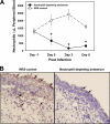
Fig. 1.
Fig. 1.
Antineutrophil antiserum treatment effectively depleted neutrophils from peripheral blood and C. caviae-infected conjunctivae of guinea pigs. Guinea pigs (n = 5/group) were administered 1.0 ml of rabbit neutrophil-depleting antiserum or the NRS control intraperitoneally every 24 h beginning the day before infection (day −1). On day 0, all animals were infected in the conjunctiva with 104 IFU of C. caviae. (A) Peripheral blood neutrophil counts were performed on guinea pigs on days −1, 0, 3, and 6 postinfection. The experiment was repeated, and values for all animals (n = 10 per treatment group) are shown as means ± standard errors of the means. *, P < 0.001 by two-way ANOVA with repeated measures (day and group) with the post hoc Holm-Sidak method for multiple comparison procedures. (B) Histological sections of C. caviae-infected conjunctivae harvested on day 4 postinfection were stained for myeloperoxidase. Red indicates myeloperoxidase/neutrophils (arrows); blue indicates nuclei of cells. Diffuse, red background staining in the NRS control represents free myeloperoxidase molecules released from neutrophils into the microenvironment. Magnification, ×200. Photomicrographs are representative of responses in each group (n = 5/group).
Fig. 2.
Fig. 2.
Neutrophil depletion did not affect the infection course (number of IFU) during the first 7 days of a primary C. caviae ocular infection. Conjunctival swabs were obtained from NRS control and neutrophil-depleted guinea pigs infected with C. caviae (104 IFU) on days 3 and 6 postinfection (n = 5 per group). IFU were isolated from swabs and measured as described in Materials and Methods. The experiment was repeated, and values for all animals (n = 10 per group) are shown as means ± standard deviations. An unpaired t test was used to statistically compare data from both experimental groups on each day. No significant differences were noted on day 3 or day 6.
Fig. 3.
Fig. 3.
Neutrophil depletion significantly decreased ocular pathology during a C. caviae ocular infection. (A) Gross ocular pathology in NRS control and neutrophil-depleted guinea pigs was visually assessed and scored on a daily basis as described in Materials and Methods. The experiment was repeated, and values for all animals (10 per group) are shown as means ± standard errors of the means (SEM). Data were analyzed using the Mann-Whitney rank sum test (*, P < 0.001). (B) H&E-stained conjunctival sections (5 μm) from NRS control and neutrophil-depleted guinea pigs on day 4 (n = 4 animals per group) and day 7 (n = 5 animals per group) were evaluated by a veterinary pathologist for mucosal epithelium erosion, based on a 0 to 4+ scaling system. Data shown are means ± SEM, and for each day the two groups were statistically compared using the Mann-Whitney rank sum test (*, P < 0.05). (C) H&E-stained sections of infected conjunctivae of guinea pigs on day 4 postinfection revealed severely damaged mucosal epithelium in NRS control guinea pigs but much less damage in neutrophil-depleted animals. A yellow bracket spans the conjunctival mucosal epithelium, showing a close-to-normal mucosal structure in neutrophil-depleted conjunctiva. Magnification, ×400. Photomicrographs are representative of responses in each group.
Fig. 4.
Fig. 4.
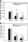
CD4+ and CD8+ T cells, but not B cells, were significantly decreased in C. caviae-infected conjunctivae in neutrophil-depleted guinea pigs. On day 7 postinfection guinea pigs were euthanized and conjunctivae harvested and processed into single-cell suspensions for immunofluorescent staining and flow cytometric analysis, as described in Materials and Methods. For the flow cytometric analysis, a gate was set on live, intact cells, using the Live/Dead cell stain kit (Invitrogen) to exclude debris and dead cells. The gated cells were then analyzed for the presence of CD4+ T cells, CD8+ T cells, and B cells. Data are presented as means ± standard deviations of the absolute number (A) and percentage (B) of lymphocyte subsets in total live cells analyzed per infected conjunctiva. Cell types in the two groups were statistically compared using an unpaired t test (*, P < 0.05; **, P < 0.005).
Fig. 5.
Fig. 5.
C. caviae-specific IgA titers were significantly increased in tears and serum with neutrophil depletion during a C. caviae ocular infection, but C. caviae-specific IgG titers in the serum were unchanged. Tears were collected on days 0, 3, and 6, and serum was obtained on days 0 and 6 postinfection from guinea pigs with C. caviae ocular infections. C. caviae-specific IgA titers were measured by an indirect ELISA and C. caviae-specific IgG titers were measured by direct ELISA as described in Materials and Methods. The experiment was repeated, and values for all animals (n = 10 per group) are shown as means ± standard deviations. Data for IgA in tears were analyzed using a two-way ANOVA with repeated measures (day and group) with the post hoc Holm-Sidak method for multiple comparison procedures. For serum, levels of IgA or IgG of both treatment groups on day 6 were compared to each other with an unpaired t test (*, P < 0.05; **, P < 0.01).
Fig. 6.
Fig. 6.
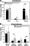
Cytokine/chemokine mRNA expression in C. caviae-infected conjunctivae was significantly altered in neutrophil-depleted guinea pigs. On day 7 postinfection, guinea pigs were euthanized and conjunctivae were harvested for quantitative PCR analysis as described in Materials and Methods. The experiment was repeated, and values for all animals (n = 10 per treatment group) are shown as means ± standard deviations. Fold increase mRNA was determined from the CT values, which were normalized for 18S rRNA expression and then normalized to the values from conjunctivae of healthy, uninfected guinea pigs. Data from the two groups were statistically compared by using an unpaired t test (*, P < 0.05; **, P < 0.02).

References

    1. Abbas A. K., Lichtman A. H., Pillai S. 2007. Cellular and molecular immunology. Saunders Elsevier, Philadelphia, PA
    1. Ahmad A., Dawson C. R., Yoneda C., Togni B., Schachter J. 1977. Resistance to reinfection with a chlamydial agent (guinea pig inclusion conjunctivitis agent). Invest. Ophthalmol. Vis. Sci. 16:549–553 - PubMed
    1. Allen S. S., McMurray D. N. 2003. Coordinate cytokine gene expression in vivo following induction of tuberculous pleurisy in guinea pigs. Infect. Immun. 71:4271–4277 - PMC - PubMed
    1. Bai H., et al. 2005. Intranasal inoculation of Chlamydia trachomatis mouse pneumonitis agent induces significant neutrophil infiltration which is not efficient in controlling the infection in mice. Immunology 114:246–254 - PMC - PubMed
    1. Barteneva N., Theodor I., Peterson E. M., de la Maza L. M. 1996. Role of neutrophils in controlling early stages of a Chlamydia trachomatis infection. Infect. Immun. 64:4830–4833 - PMC - PubMed

Publication types