. 2011 May;31(10):2111-21.
doi: 10.1128/MCB.01159-10. Epub 2011 Mar 14.

Importin beta plays an essential role in the regulation of the LysRS-Ap(4)A pathway in immunologically activated mast cells

Affiliations

Importin beta plays an essential role in the regulation of the LysRS-Ap(4)A pathway in immunologically activated mast cells

Irit Carmi-Levy et al. Mol Cell Biol. 2011 May.

Abstract

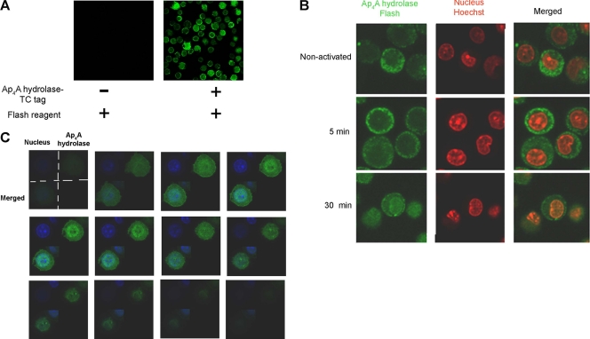
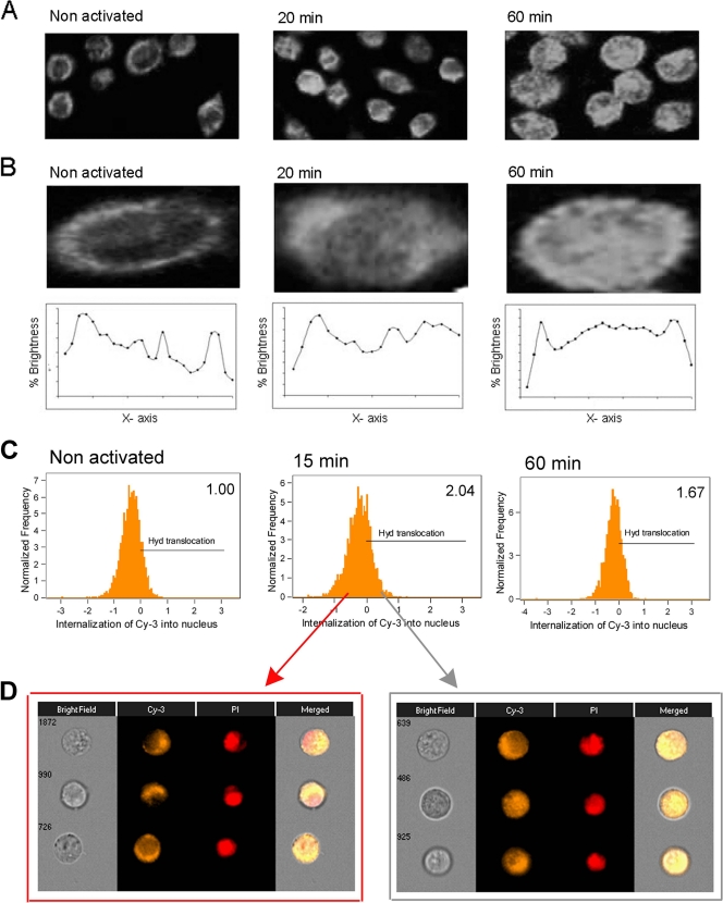
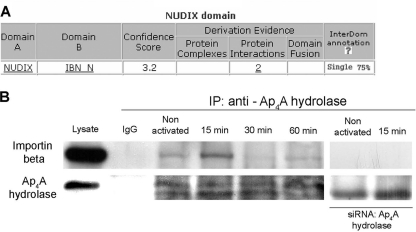
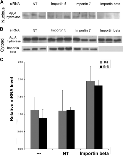
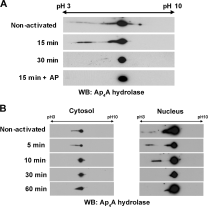
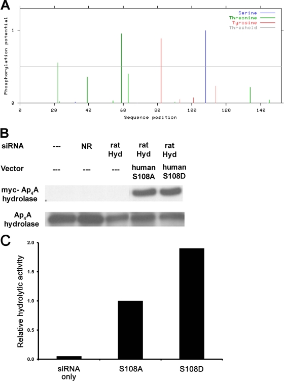
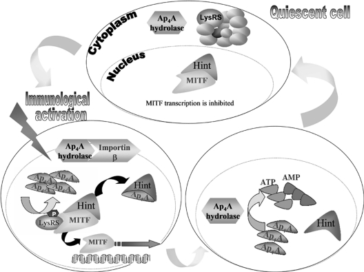
We recently reported that diadenosine tetraphosphate hydrolase (Ap(4)A hydrolase) plays a critical role in gene expression via regulation of intracellular Ap(4)A levels. This enzyme serves as a component of our newly described lysyl tRNA synthetase (LysRS)-Ap(4)A biochemical pathway that is triggered upon immunological challenge. Here we explored the mechanism of this enzyme's translocation into the nucleus and found its immunologically dependent association with importin beta. Silencing of importin beta prevented Ap(4)A hydrolase nuclear translocation and affected the local concentration of Ap(4)A, which led to an increase in microphthalmia transcription factor (MITF) transcriptional activity. Furthermore, immunological activation of mast cells resulted in dephosphorylation of Ap(4)A hydrolase, which changed the hydrolytic activity of the enzyme.


Figures

Fig. 1.
Fig. 1.
Ap4A hydrolase translocates into the nuclei of immunologically activated BMMC. (A) BMMC were transfected with an Ap4A hydrolase-TC tag-expressing vector. Transfected cells exhibited fluorescence at the predicted wavelength, while no fluorescence was detected in nontransfected cells. (B) BMMC were transfected with Ap4A hydrolase-TC tag. The cells were immunologically activated for specific times, plated on chambered coverslips, and labeled with 2 μM FlAsH. The cell nuclei were stained with Hoechst stain. Confocal microscopy was used to evaluate Ap4A hydrolase subcellular localization. One representative field of three is presented. (C) BMMC were transfected with Ap4A hydrolase-TC tag and activated for 30 min. z-Stack analysis of Ap4A hydrolase was performed using confocal microscopy to obtain sequential z-axis images.
Fig. 2.
Fig. 2.
Identification of Ap4A hydrolase nuclear translocation in RBL cells following IgE-Ag activation. (A) RBL cells were either quiescent or sensitized with IgE and then were challenged with antigen for 20 or 60 min. Immunostaining was performed with anti-Ap4A hydrolase. The cells were analyzed by confocal laser scanning microscopy. Images for one representative experiment of three are presented. (B) One representative cell from the field is shown at high magnification. Using Photoshop picture-editing software, the percent brightness parameter was determined at specific intervals along the x axis. (C) RBL cells were activated (or not) with IgE, challenged with Ag for specific times, and then stained for Ap4A hydrolase by anti-Ap4A hydrolase followed by Cy3-conjugated anti-rabbit, together with PI staining to visualize nuclei. Nuclear translocation was evaluated by ImageStream flow cytometry. For each treatment, 5,000 cells were collected. Positive single cells were gated, and nuclear translocation of Ap4A hydrolase was plotted. The relative translocated population size is indicated in the upper right corner. (D) Representative images of untranslocated (left) and translocated (right) Ap4A hydrolase (Cy3 column), as well as PI and merged images, for the 15-min treatment.
Fig. 3.
Fig. 3.
Substantial association between Ap4A hydrolase and importin beta in RBL cells. (A) The protein sequence of Ap4A hydrolase was subject to domain-domain association prediction analysis using bioinformatic tools. The Nudix domain was predicted to associate with the importin beta N-terminal domain (IBN_N) by the putative interaction database InterDom (http://interdom.i2r.a-star.edu.sg/). (B) Coimmunoprecipitation analysis demonstrating the time course of Ap4A hydrolase-importin beta interaction following IgE-Ag stimulation, using anti-Ap4A hydrolase antibody for immunoprecipitation (IP) and either anti-importin beta or anti-Ap4A hydrolase for immunoblotting. The specific Ap4A hydrolase band was verified by a control experiment using siRNA to knock down Ap4A hydrolase (right lanes). Data for one representative experiment of six are shown.
Fig. 4.
Fig. 4.
Importin beta-mediated Ap4A hydrolase nuclear translocation is involved in MITF transcriptional activity regulation. (A and B) RBL cells were transfected with siRNA oligonucleotides against importin beta, 5, and 7. NT siRNA was used as a control. Nuclear fractions were isolated from cells activated with IgE and antigen for 30 min. Nuclear extracts (A) and cytosolic extracts (B) were analyzed by Western blotting with anti-Ap4A hydrolase antibody. Silencing of importin beta was verified by Western blotting with anti-importin beta. Each lane represents an independent experiment. (C) RBL cells were transfected with either importin beta siRNA or NT siRNA. Control and transfected cells were activated with IgE-Ag. Twenty-four hours later, cells were lysed and mRNA quantitation for GrB and Kit was performed by SYBR green incorporation into real-time PCR with RBL cells. Expression levels were normalized to those of the β-actin housekeeping gene. Results are presented as relative quantities, with the untreated cell level arbitrarily set to 1. The means and standard errors of the means for three experiments are shown.
Fig. 5.
Fig. 5.
Ap4A hydrolase is subject to dephosphorylation upon IgE-Ag activation. (A) Lysates from IgE-Ag-activated and nonactivated RBL cells were subjected to 2D electrophoresis on a pH 3 to 10 gradient in an 8% polyacrylamide gel. The gel was blotted with anti-Ap4A hydrolase antibody. WB, Western blot. (B) Nuclear and cytoplasmic fractions were isolated from RBL cells activated with IgE and antigen. The subcellular extracts were analyzed by 2D electrophoresis with anti-Ap4A hydrolase antibody.
Fig. 6.
Fig. 6.
Phosphorylation status of serine 108 in Ap4A hydrolase is involved in the enzymatic activity of Ap4A metabolism. (A) Putative serine, threonine, and tyrosine phosphorylation sites in the Ap4A hydrolase protein sequence, as predicted by the NetPhos 2.0 program (www.cbs.dtu.dk/services/NetPhos/). (B) RBL cells were transfected with rat Ap4A hydrolase siRNA. Twenty-four hours later, cells were transfected with human Ap4A hydrolase S108A and S108D variants. Next, the cells were incubated with IgE and challenged with DNP for 30 min. The cell extracts were analyzed by Western blotting with anti-Myc or anti-Ap4A hydrolase antibodies. (C) RBL cell lysates from panel B were subjected to an enzymatic activity assay. Ap4A hydrolase hydrolytic activity was determined as the rate of synthetic Ap4A degradation, which was evaluated by chemiluminescence detection of ATP as described in Materials and Methods. Data for one representative experiment of three are shown.
Fig. 7.
Fig. 7.
Proposed model for involvement of Ap4A hydrolase in transcription regulation. Upon immunological activation, Ap4A hydrolase associates with importin beta and is translocated into the nucleus when Ap4A levels are elevated. Ap4A binds to Hint-1, dissociating it from MITF. With the repression removed, transcription factors are free to transcribe their target genes. The presence of Ap4A hydrolase in the nucleus leads to hydrolysis of the accumulated Ap4A into AMP and ATP, decreasing its levels to that found in resting cells. When Ap4A levels decrease, Hint-1 reassociates with MITF.

References

    1. Abdelghany H. M., et al. 2001. Cloning, characterisation and crystallisation of a diadenosine 5′, 5‴-P(1),P(4)-tetraphosphate pyrophosphohydrolase from Caenorhabditis elegans. Biochim. Biophys. Acta 1550:27–36 - PubMed
    1. Bayliss R., Littlewood T., Strawn L. A., Wente S. R., Stewart M. 2002. GLFG and FxFG nucleoporins bind to overlapping sites on importin-beta. J. Biol. Chem. 277:50597–50606 - PubMed
    1. Blom N., Gammeltoft S., Brunak S. 1999. Sequence and structure-based prediction of eukaryotic protein phosphorylation sites. J. Mol. Biol. 294:1351–1362 - PubMed
    1. Bousset K., Diffley J. F. 1998. The Cdc7 protein kinase is required for origin firing during S phase. Genes Dev. 12:480–490 - PMC - PubMed
    1. Reference deleted.

Publication types

MeSH terms

Substances

LinkOut - more resources