. 2011 May;31(10):2162-70.
doi: 10.1128/MCB.05234-11. Epub 2011 Mar 14.

Podocyte-specific deletion of Myh9 encoding nonmuscle myosin heavy chain 2A predisposes mice to glomerulopathy

Affiliations

Podocyte-specific deletion of Myh9 encoding nonmuscle myosin heavy chain 2A predisposes mice to glomerulopathy

Duncan B Johnstone et al. Mol Cell Biol. 2011 May.

Abstract

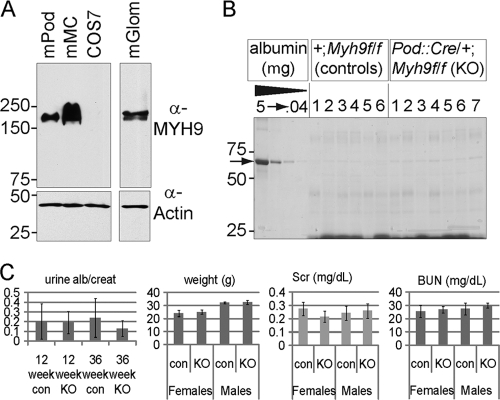
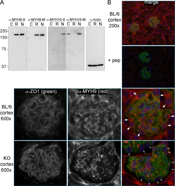
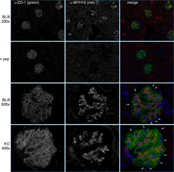
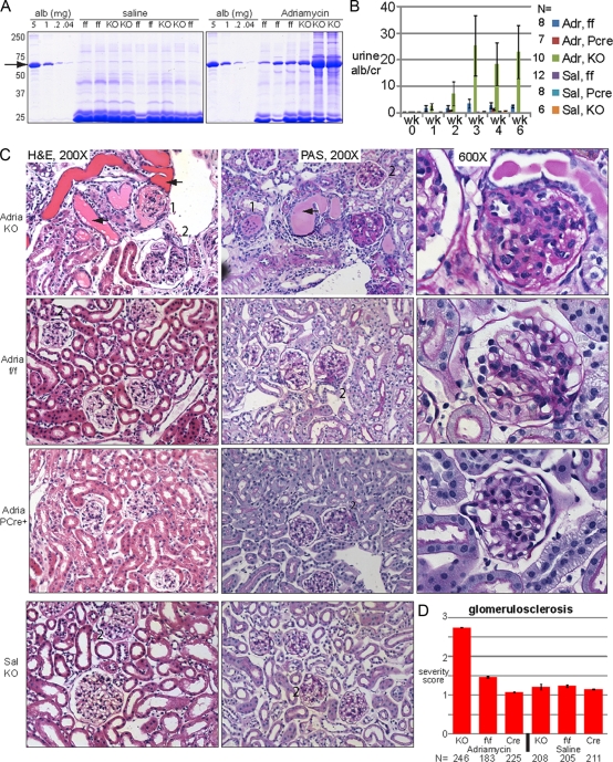
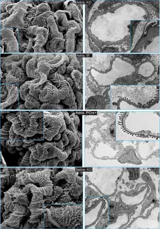
Genome-wide association studies linked single-nucleotide polymorphisms (SNPs) at the MYH9 locus to chronic kidney disease among African-Americans, particularly glomerular diseases such as HIV nephropathy and idiopathic focal and segmental glomerulosclerosis (FSGS). However, these MYH9 SNPs are intronic, and despite extensive sequencing, a causal variant remains elusive. To investigate the role of MYH9 in kidney disease, we selectively deleted Myh9 from mouse podocytes and found that mutant C57BL/6 mice did not develop renal insufficiency or proteinuria compared to control littermates, even when the mice were aged for 9 months. To explain the surprisingly normal phenotype, we considered genetic redundancy with the paralog Myh10 in podocytes, but we found that Myh10 was not expressed in podocytes in Myh9-deficient or control mice. We tested whether Myh9 podocyte deletion predisposed mice to glomerulopathy in response to injury by doxorubicin hydrochloride (Adriamycin), and we found that Myh9 podocyte-deleted mice developed proteinuria and glomerulosclerosis, while control mice were resistant. In summary, Myh9 podocyte deletion in C57BL/6 mice results in susceptibility to experimental doxorubicin hydrochloride glomerulopathy. We review evidence that MYH9 dysfunction in humans results in similar susceptibility and place our data, the first examination of Myh9 kidney disease in experimental animals, in the context of recent findings in human kidney disease, including the role of APOL1.


Figures

Fig. 1.
Fig. 1.
Initial characterization of MYH9 in the murine kidney. (A) MYH9 immunoblot of lysates separated by SDS-PAGE. The lanes are for mouse podocytes (mPod), mouse mesangial cells (mMC), COS7 cells (which do not express MYH9), and mouse glomerular lysate (mGlom). (B) Screen for proteinuria in adult mouse urine. Shown is one of two gels with urines from 4-month-old mice of the indicated genotype, separated by SDS-PAGE and stained with Coomassie blue. Each lane is for the urine from one mouse, with a BSA standard shown on the left, in 5-fold dilutions. The arrow indicates albumin. For all urine samples, low-molecular-mass proteins of 20 to 25 kDa are visible, but albumin is barely visible (0.04 μg or less). (C) Phenotype of podocyte-specific KO mice. The left graph shows urine albumin/creatinine ratios for 12- and 36-week-old mice. In the next three graphs, for weights and serum creatinine (Scr) and blood urea nitrogen (BUN) levels of 9-month-old mice, there is no significant difference between KO and control mice (all P values are >0.1 by t test).
Fig. 2.
Fig. 2.
Localization of MYH9 in mouse kidneys. (A) Immunoblots to characterize new anti-MYH9 and anti-MYH10 antibodies, comparing equivalent titers of crude serum (S) or Melon gel-purified antibody (M). Lysates: C, COS7 cells (which do not express MYH9); R, rat basophil leukemia cells (which do not express MYH10); and N, NIH 3T3 cells (which express both MYH9 and MYH10). (B) Indirect IF with purified anti-MYH9 (1:400) and anti-ZO1 (1:200) in perfusion-fixed kidneys. (Top right) The panel (merge) is a ×200 view demonstrating ZO1 in glomeruli and MYH9 in a subset of tubules as well as glomeruli. (Top right, second panel) MYH9 peptide (1 ng/μl) was added to the anti-MYH9 and anti-ZO1 primary Ab mix (+ pep), and with identical photo settings, there was no MYH9 staining, while ZO1 staining was unchanged. Secondary Ab-only controls were similarly dark. (Top row) A ×600 view of a glomerulus demonstrates anti-MYH9 staining of podocyte cytoplasm (large white arrows) as well as mesangial and possibly endothelial cells within the glomerulus. (Bottom row) In glomeruli from KO mice, MYH9 is no longer visible in podocytes (small white arrows), while MYH9 staining of the mesangium and other cells is unchanged.
Fig. 3.
Fig. 3.
Localization of MYH10 in mouse kidneys. Indirect IF was performed with anti-MYH10 (1:800), as characterized by immunoblotting in Fig. 2A, and anti-ZO1 (1:200). (Top row) Images at a magnification of ×200 show MYH10 in glomeruli and rare tubules. (Second row) MYH10 peptide (0.2 ng/μl) was added to the anti-MYH10 and anti-ZO1 primary Ab mix (+ pep). With identical photo settings, MYH10 staining was competed away while ZO1 staining was unchanged. (Third row) Images of glomeruli at a magnification of ×600 reveal MYH10 in mesangial cells but not in podocytes (small white arrows). (Bottom row) MYH10 expression in kidneys of KO mice is unchanged from that in controls, with prominent mesangial staining and no staining of podocytes (small white arrows).
Fig. 4.
Fig. 4.
Podocyte-specific KO mice are predisposed to doxorubicin hydrochloride-induced nephropathy. KO, knockout mice (Pod::Cre/+; Myh9f/f); ff, control mice (+; Myh9f/f); PCre, control mice (PCre; +/+); Adr, doxorubicin hydrochloride (Adriamycin) in saline; Sal, saline only. (A) Coomassie blue-stained SDS-PAGE gel with urines 2 weeks after injection of saline or doxorubicin hydrochloride. An albumin standard is shown at 5-fold dilutions, with the arrow indicating albumin. All urines with heavy albuminuria were from KO animals. (B) Urine ELISA albumin/creatinine ratios after injection of mice with saline or doxorubicin hydrochloride (means ± standard errors of the means). Doxorubicin hydrochloride-treated KO mice had more albuminuria than all other groups at weeks 3, 4, and 6 (Welsch's t test [unpaired, not assuming equal variances, and one-tailed]). All pairwise combinations with doxorubicin hydrochloride-treated KO mice had P values of 0.05 or less, although due to the large standard error of the mean, the significance was borderline for doxorubicin hydrochloride-treated KO versus doxorubicin hydrochloride-treated ff mice at 4 weeks (P value = 0.0524) and for doxorubicin hydrochloride-treated KO versus doxorubicin hydrochloride-treated ff mice at 3 weeks (P value = 0.0526). (C) KO mice develop focal and segmental glomerulosclerosis. Some images were removed to accommodate available space. 1, global sclerosis; 2, segmental sclerosis; arrow, proteinaceous cast. Images for ff control mice and PCre+ mice injected with saline were similar to those for KO mice injected with saline and are not shown. (D) Mean glomerulosclerosis severity scores (± standard deviations), judged blindly, albeit subjectively (1 to 4+), for two mice for each condition. N, total number of glomeruli. Median scores are reported in Results. By the nonparametric Mann-Whitney test, there was no difference between saline-treated groups or between PCre controls treated with doxorubicin hydrochloride versus saline, but there was a significant difference between KO mice treated with doxorubicin hydrochloride and every other group (P < 1e−6).
Fig. 5.
Fig. 5.
Scanning electron microscopy (left panels) and transmission electron microscopy (right panels) reveal changes in foot process architecture in KO mice injected with doxorubicin hydrochloride. Scanning EM of glomeruli was performed at low power (×3,000) and high power (inset; ×10,000). Transmission EM images were obtained at low power (×6,000 to ×7,500) and high power (inset; ×15,000 to ×25,000). Genotypes are indicated in the center of each row. Images for ff control mice and PCre+ control mice injected with saline were similar to those for KO mice injected with saline and are not shown.

References

    1. Arrondel C., et al. 2002. Expression of the nonmuscle myosin heavy chain IIA in the human kidney and screening for MYH9 mutations in Epstein and Fechtner syndromes. J. Am. Soc. Nephrol. 13:65–74 - PubMed
    1. Bao J., Jana S. S., Adelstein R. S. 2005. Vertebrate nonmuscle myosin II isoforms rescue small interfering RNA-induced defects in COS-7 cell cytokinesis. J. Biol. Chem. 280:19594–19599 - PubMed
    1. Behar D. M., et al. 2010. African ancestry allelic variation at the MYH9 gene contributes to increased susceptibility to non-diabetic end-stage kidney disease in Hispanic Americans. Hum. Mol. Genet. 19:1816–1827 - PMC - PubMed
    1. Conti M. A., Adelstein R. S. 2008. Nonmuscle myosin II moves in new directions. J. Cell Sci. 121:11–18 - PubMed
    1. Conti M. A., Even-Ram S., Liu C., Yamada K. M., Adelstein R. S. 2004. Defects in cell adhesion and the visceral endoderm following ablation of nonmuscle myosin heavy chain II-A in mice. J. Biol. Chem. 279:41263–41266 - PubMed

Publication types

MeSH terms