. 2011 Apr 15;186(8):4546-50.
doi: 10.4049/jimmunol.1004021. Epub 2011 Mar 14.

Cutting edge: Generation of colitogenic Th17 CD4 T cells is enhanced by IL-17+ γδ T cells

Affiliations

Cutting edge: Generation of colitogenic Th17 CD4 T cells is enhanced by IL-17+ γδ T cells

Jeong-su Do et al. J Immunol. .

Abstract

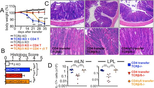
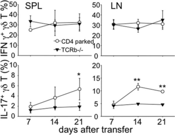
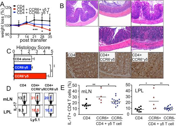
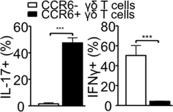
Th 17 cells have been implicated in the pathogenesis of colitis; however, a cellular mechanism by which colitogenic Th17 immunity arises in vivo remains unclear. In this study, we report that a subset of IL-17(+) γδ T cells plays a crucial role in enhancing in vivo Th17 differentiation and T cell-mediated colitis. TCRβ(-/-) mice were highly susceptible to T cell-mediated colitis, whereas TCRβδ(-/-) mice were resistant to the disease. Importantly, cotransfer of IL-17(+) but not of IL-17(-) γδ T cells with CD4 T cells was sufficient to enhance Th17 differentiation and induce full-blown colitis in TCRβδ(-/-) recipients. Collectively, our results provide a novel function of IL-17(+) γδ T cell subsets in supporting in vivo Th17 differentiation and possibly in fostering the development of intestinal inflammation.


Figures

Figure 1
Figure 1. TCRβ-/- recipients of naïve CD4 T cells develop exacerbated colitis while TCRβδ-/- recipients are fully resistant to the colitis, and adoptive transfer of γδ T cells restores colitis in TCRβδ-/- recipients
(A) FACS purified CD4+CD25-CD44low Thy1.1 naïve T cells (2.5 × 10⁵) were transferred into TCRβ-/- or TCRβδ-/- mice. In some experiments, FACS sorted γδ T cells (2.5 × 10⁵) were cotransferred with CD4 T cells into TCRβδ-/- mice. Body weight was weekly monitored and presented as a percentage of the initial weight at day 0. ** p<0.01; *** p <0.001. Data shown are from 5-8 individually tested mice. (B) Colitis score of each group. (C) Representative H&E staining of colon 5 weeks post transfer. 20× original magnification. (D) Total donor cell recovery of IL-17+ cells were calculated. Each symbol represents individually tested mouse. *** p<0.001.
Figure 2
Figure 2. Increased IL-17+ γδ T cells after CD4 T cell transfer
TCRβ-/- mice were transferred with naïve CD4 T cells as described above. Spleen and LN cells were harvested every week post transfer and ex vivo stimulated and γδ T cell cytokine expression was examined. The results shown represent the mean ± SD of 4-5 individually tested mice. * p<0.05; ** p<0.01.
Figure 3
Figure 3. CCR6+ γδ T cells support Th17 differentiation and colitogenicity of CD4 T cells in vivo
CD4+CD25-CD44low Ly5.1 naïve T cells (2.5 × 10⁵) were transferred into TCRβδ-/- recipients with sorted CCR6- γδ T cells or CCR6+ γδ T cells (2.5 × 10⁵). (A) Weight was weekly monitored. * p<0.05; *** p<0.001 (compared with CD4 alone group). (B) Representative H&E staining and immunohistochemistry for CD3 staining of colon 5 weeks post transfer of CD4 alone, CD4/CCR6- γδ T cell co-transfer, and CD4/CCR6+ γδ T cell co-transfer. 20X original magnification. (C) Colitis score of each group. (D and E) mLN and LP cells were isolated 5 weeks post transfer. The percentage of IL-17 producing Ly5.1 CD4 T cells from TCRβδ-/- recipients of CD4 alone, CD4/CCR6- γδ T cell co-transfer, and CD4/CCR6+ γδ T cell cotransfer is shown. Each symbol represents individually tested mouse. * p<0.05; ** p<0.01.
Figure 4
Figure 4. Cytokine profiles of γδ T cells are stable in vivo
Peripheral LN γδ T cells from Ly5.1 mice were sorted based on CCR6 expression and separately transferred into Rag2-/- mice (2.5×10⁵). Spleen cells were harvested 14 days post transfer and ex vivo stimulated for cytokine expression. Shown are IFNγ and IL-17 expression of Ly5.1 gated γδ T cells. The results shown represent the mean ± SD from two independent experiments.

References

    1. Zhu J, Yamane H, Paul WE. Differentiation of effector CD4 T cell populations (*). Annu Rev Immunol. 2010;28:445–489. - PMC - PubMed
    1. Carding SR, Egan PJ. Gammadelta T cells: functional plasticity and heterogeneity. Nat Rev Immunol. 2002;2:336–345. - PubMed
    1. Cua DJ, Tato CM. Innate IL-17-producing cells: the sentinels of the immune system. Nat Rev Immunol. 2010;10:479–489. - PubMed
    1. Roark CL, Simonian PL, Fontenot AP, Born WK, O'Brien RL. gammadelta T cells: an important source of IL-17. Curr Opin Immunol. 2008;20:353–357. - PMC - PubMed
    1. Do JS, Fink PJ, Li L, Spolski R, Robinson J, Leonard WJ, Letterio JJ, Min B. Cutting edge: spontaneous development of IL-17-producing gamma delta T cells in the thymus occurs via a TGF-beta 1-dependent mechanism. J Immunol. 2010;184:1675–1679. - PMC - PubMed

Publication types

MeSH terms

Substances