. 2011 Mar 29;108(13):5354-9.
doi: 10.1073/pnas.1019378108. Epub 2011 Mar 14.

Microbiota regulates immune defense against respiratory tract influenza A virus infection

Affiliations

Microbiota regulates immune defense against respiratory tract influenza A virus infection

Takeshi Ichinohe et al. Proc Natl Acad Sci U S A. .

Abstract

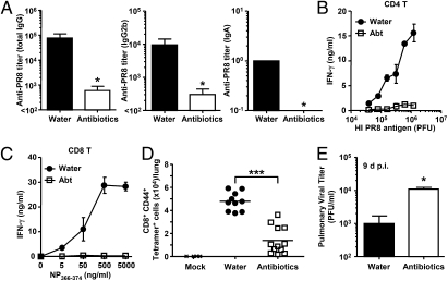
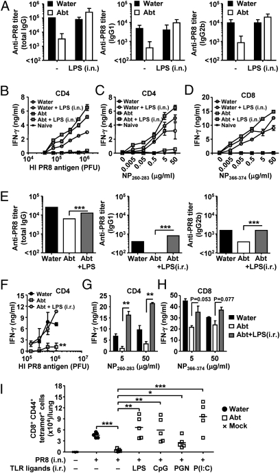
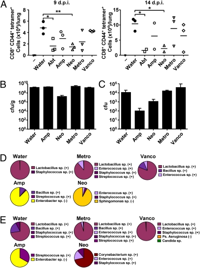
Although commensal bacteria are crucial in maintaining immune homeostasis of the intestine, the role of commensal bacteria in immune responses at other mucosal surfaces remains less clear. Here, we show that commensal microbiota composition critically regulates the generation of virus-specific CD4 and CD8 T cells and antibody responses following respiratory influenza virus infection. By using various antibiotic treatments, we found that neomycin-sensitive bacteria are associated with the induction of productive immune responses in the lung. Local or distal injection of Toll-like receptor (TLR) ligands could rescue the immune impairment in the antibiotic-treated mice. Intact microbiota provided signals leading to the expression of mRNA for pro-IL-1β and pro-IL-18 at steady state. Following influenza virus infection, inflammasome activation led to migration of dendritic cells (DCs) from the lung to the draining lymph node and T-cell priming. Our results reveal the importance of commensal microbiota in regulating immunity in the respiratory mucosa through the proper activation of inflammasomes.


Conflict of interest statement

The authors declare no conflict of interest.

Figures

Fig. 1.
Fig. 1.
Antibiotic-treated mice fail to induce acquired immunity to influenza virus infection. C57BL/6 mice were given ampicillin (1 g/L), vancomycin (500 mg/L), neomycin sulfate (1 g/L), and metronidazole (1 g/L) in drinking water for 4 wk before PR8 virus infection (10 pfu per mouse). Two weeks later, serum and nasal wash were collected and Ag-specific antibody titers were measured (A), and T-cells were isolated from spleen and restimulated with flu virion or NP peptide for 72 h, and IFN-γ production from CD4 T cells (B) and CD8 T cells (C) was measured. (D) Lymphocytes were collected from the lung of infected animals at 14 d postinfection and stained with flu-specific tetramer. (E) The lung washes of flu-infected mice were harvested at 9 d postinfection, and viral titers were measured by plaque assay. *P < 0.05 and ***P < 0.001 vs. water-fed group. Data represent the mean ± SD. Similar results were obtained from three separate experiments.
Fig. 2.
Fig. 2.
Local and distal TLR stimulation restores immune response to influenza virus infection in antibiotic-treated mice. C57BL/6 mice were given antibiotics in drinking water for 4 wk before 10 pfu of PR8 viral infection with or without 2 μg of LPS injected intranasally (AD) or intrarectally (EH). Two weeks later, serum was collected and Ag-specific antibody titers were measured (A and E), and T cells were isolated from spleen and restimulated with flu virion (B and F) or NP peptide (C, D, G, and H) for 72 h, and IFN-γ production from CD4 T cells (B, C, F, and G) and CD8 T cells (D and H) was measured. (I) Water-fed and antibiotic-treated mice were infected intranasally with 10 pfu of PR8 virus with or without intrarectal injection of LPS (5 μg), CpG2216 (50 μg), peptidoglycan (20 μg), or Poly (I:C) (50 μg). Total numbers of influenza virus-specific CD8 T cells in the lung are shown. Data represent the mean ± SD, and are representative of at least three independent experiments (AH) or are pooled from two independent experiments (I). *P < 0.05; **P < 0.01; ***P < 0.001.
Fig. 3.
Fig. 3.
Effect of single antibiotic treatment on bacterial colonization and immune responses to respiratory influenza virus infection. C57BL/6 mice were each given single or four combinatorial antibiotics in drinking water for 4 wk before PR8 virus infection (10 pfu per mouse). Lymphocytes were collected from the lung of infected animals at 9 d or 14 d postinfection and stained with flu-specific tetramer (A). Bacterial load in the stool (B) and nasal wash (C) from antibiotic-treated mice (n = 3 per condition) were measured. Bacterial compositions in the stool (D) and nasal wash (E) from single antibiotic-treated mice are depicted. Purple and yellow tones denote Gram-positive and Gram-negative bacteria, respectively. Similar results were obtained from two to three separate experiments. *P < 0.05; **P < 0.01.
Fig. 4.
Fig. 4.
Respiratory tract DCs fail to migrate to the draining LN and prime T-cell responses in antibiotic-treated mice. C57BL/6 mice were given antibiotics in drinking water for 4 wk before intranasal infection with 1,000 pfu of PR8-GP33 viruses. (AC) Three days later, CD11c+ DCs were isolated from the mediastinal LN. (DF) Naïve p14 tg CD8 T cells (2 × 10⁵ cells per well) were cocultured with different numbers of CD11c+ DCs isolated from mLN of infected animals with (F) or without GP33 peptide (D and E) for 72 h. Splenic DCs from infected animals were used as negative control (E). IFN-γ production (E and F) and the number of CD8 T cells (D) were measured. The number of CD103+ DCs (G) and phenotype of total DC population (H) in the lung and mLN were measured in antibiotic-treated mice without influenza infection. (I and J) Water-fed, antibiotic-treated, and caspase-1–deficient mice were inoculated intranasally with CFSE. Six hours later, mice were infected with 1,000 pfu of PR8 viruses. Eighteen hours after infection, mediastinal LNs were collected. The numbers of CFSE+CD11c+DCs are shown. (K) LPS inoculation (intranasal or intrarectal) restored DC migration to the mLN following intranasal influenza virus infection. Data represent the mean ± SD. Similar results were obtained from three separate experiments.

Comment in

References

    1. Neumann G, Noda T, Kawaoka Y. Emergence and pandemic potential of swine-origin H1N1 influenza virus. Nature. 2009;459:931–939. - PMC - PubMed
    1. Fitzgerald KA. NLR-containing inflammasomes: Central mediators of host defense and inflammation. Eur J Immunol. 2010;40:595–598. - PubMed
    1. Schmitz N, Kurrer M, Bachmann MF, Kopf M. Interleukin-1 is responsible for acute lung immunopathology but increases survival of respiratory influenza virus infection. J Virol. 2005;79:6441–6448. - PMC - PubMed
    1. Allen IC, et al. The NLRP3 inflammasome mediates in vivo innate immunity to influenza A virus through recognition of viral RNA. Immunity. 2009;30:556–565. - PMC - PubMed
    1. Thomas PG, et al. The intracellular sensor NLRP3 mediates key innate and healing responses to influenza A virus via the regulation of caspase-1. Immunity. 2009;30:566–575. - PMC - PubMed

Publication types

MeSH terms