. 2011 Mar 29;108(13):5402-7.
doi: 10.1073/pnas.1100410108. Epub 2011 Mar 14.

Iron enzyme ribulose-5-phosphate 3-epimerase in Escherichia coli is rapidly damaged by hydrogen peroxide but can be protected by manganese

Affiliations

Iron enzyme ribulose-5-phosphate 3-epimerase in Escherichia coli is rapidly damaged by hydrogen peroxide but can be protected by manganese

Jason M Sobota et al. Proc Natl Acad Sci U S A. .

Abstract

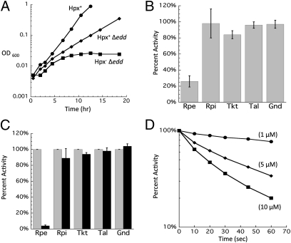
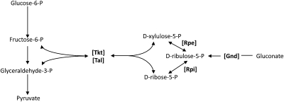
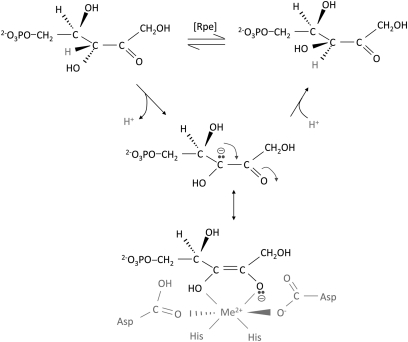
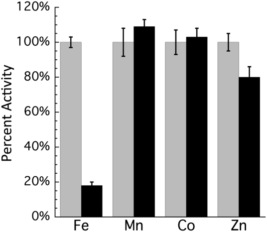
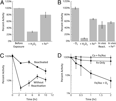
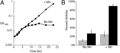
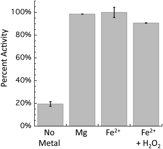
H(2)O(2) is commonly generated in biological habitats by environmental chemistry and by cellular immune responses. H(2)O(2) penetrates cells, disrupts metabolism, and blocks growth; it therefore is of interest to identify the major cellular molecules that H(2)O(2) damages and the strategies by which cells protect themselves from it. We used a strain of Escherichia coli that lacks catalases and peroxidases to impose protracted low-grade H(2)O(2) stress. Physiological analysis indicated that the pentose-phosphate pathway, in particular, was poisoned by submicromolar intracellular H(2)O(2). Assays determined that ribulose-5-phosphate 3-epimerase (Rpe) was specifically inactivated. In vitro studies demonstrated that Rpe employs a ferrous iron atom as a solvent-exposed cofactor and that H(2)O(2) rapidly oxidizes this metal in a Fenton reaction. The oxidized iron is released immediately, causing a loss of activity. Most Rpe proteins could be reactivated by remetallation; however, a small fraction of proteins were irreversibly damaged by each oxidation cycle, and so repeated cycles of oxidation and remetallation progressively led to permanent inactivation of the entire Rpe pool. Manganese import and iron sequestration are key elements of the H(2)O(2) stress response, and we found that manganese can activate Rpe in vitro in place of iron, converting the enzyme to a form that is unaffected by H(2)O(2). Indeed, the provision of manganese to H(2)O(2)-stressed cells protected Rpe and enabled the pentose-phosphate pathway to retain function. These data indicate that mononuclear iron enzymes can be primary targets of H(2)O(2) stress and that cells adapt by shifting from iron- to manganese-centered metabolism.


Conflict of interest statement

The authors declare no conflict of interest.

Figures

Fig. 1.
Fig. 1.
(A) Hpx Δedd cells cannot grow on gluconate because of damage to Rpe. After anaerobic preculture, cells were diluted into aerobic minimal gluconate medium at time 0. Data presented are representative of multiple experiments. (B) Activities measured in extracts prepared from Hpx Δedd cells grown aerobically. Values were normalized to those of anaerobic cells. Gnd, 6-phosphogluconate dehydrogenase; Rpi, ribose-5-phosphate isomerase; Tal, transaldolase; Tkt, transketolase. (C) Activities measured before (gray bars) and after (black bars) 10 μM H2O2 was added to cell extracts prepared from anaerobically grown Hpx cells. Error bars represent SD from the mean of three independent experiments. (D) Time course of the inactivation at 0 °C of Rpe in cell extracts treated with the indicated concentrations of H2O2. Extracts were prepared from anaerobically grown Hpx cells.
Fig. 2.
Fig. 2.
The pentose–phosphate pathway. Relevant intermediates are listed, with key enzymes denoted in brackets. Arrows indicate direction of each reaction.
Fig. 3.
Fig. 3.
Rpe active site structure and mechanism. Rpe interconverts ribulose-5-phosphate and xylulose-5-phosphate by abstracting a proton from one face of the C-3 carbon and then adding a proton to the opposite face. The metal coordinates substrate and stabilizes the intermediate oxyanion (25).
Fig. 4.
Fig. 4.
Sensitivity of Rpe metalloforms to H2O2 in vitro. Pure Rpe was metallated with the indicated metals. Activity then was measured before (gray bars) and after (black bars) treatment with 500 μM H2O2 for 2 min. Error bars represent SD from the mean of three independent measurements.
Fig. 5.
Fig. 5.
Reversibility of Rpe inactivation. (A) A single cycle of in vitro damage is mostly reversible. Pure Rpe was metallated with 100 μM Fe2+ before being treated with 200 μM H2O2 for 5 min. Catalase and DTPA were added to remove Fe and H2O2, and Rpe was assayed. Damaged enzyme then was reactivated by anaerobic incubation with 100 μM Fe2+ and 500 μM ascorbate. (B) Brief damage in vivo is mostly reversible. Hpx cultures were grown in anaerobic medium, and chloramphenicol then was added to block further protein synthesis. Cells then were aerated and exposed to H2O2 (100 μM) for 10 min. Cultures were returned to the anaerobic chamber, treated with catalase, and assayed before (+H2O2) or after (+Fe2+) in vitro reactivation. In vivo reactivation was measured by incubating cells for an additional 20 min after the termination of H2O2 stress. (C) Protracted stress irreversibly damages Rpe in vivo. Anaerobic cells were diluted into aerobic minimal gluconate medium at time 0. At indicated time points Rpe activity was measured before and after in vitro reactivation with Co2+. (D) Repetitive Fenton chemistry irreversibly damages Rpe in vitro. Pure Rpe was metallated with Fe2+ or Co2+. After 10 min, samples were diluted into aerobic buffer containing Fe2+ and ascorbate to trigger cycles of H2O2 damage and remetallation by Fe2+. At indicated time points, aliquots were removed, damage was terminated by the addition of catalase and DTPA, and Co2+ was added to activate the remaining functional apoenzyme. Error bars represent the SD from the mean of three experiments. The enzyme is not inactivated if the active site is occupied by Co.
Fig. 6.
Fig. 6.
Mn supplements protect Rpe against irreversible damage in vivo. (A) After anaerobic preculture, cells were diluted at time 0 into aerobic minimal gluconate medium with or without 50 μM MnCl2. Data are representative of multiple growth experiments. (B) Cells were handled as in A except that aerobic inoculum was to 0.05 OD. At various times, aliquots were taken, and Rpe activity was measured before and after reactivation with Co. Time 0 is an anaerobic time point. Error bars represent the SD from the mean of three independent experiments.
Fig. 7.
Fig. 7.
Transketolase and necessity of solvent exposure for metal sensitivity to chelation and to H2O2. Metals were added to pure transketolase (Sigma) before the addition of TPP and 500 μM H2O2. After mixing, samples were incubated for 5 min and then assayed as described in SI Materials and Methods.

References

    1. Loew O. A new enzyme of general occurrence in organisms. Science. 1900;11:701–702. - PubMed
    1. Glass GA, et al. The respiratory burst oxidase of human neutrophils. Further studies of the purified enzyme. J Biol Chem. 1986;261:13247–13251. - PubMed
    1. Mehdy MC. Active oxygen species in plant defense against pathogens. Plant Physiol. 1994;105:467–472. - PMC - PubMed
    1. Carmel-Harel O, Storz G. Roles of the glutathione- and thioredoxin-dependent reduction systems in the Escherichia coli and Saccharomyces cerevisiae responses to oxidative stress. Annu Rev Microbiol. 2000;54:439–461. - PubMed
    1. Lee JW, Helmann JD. The PerR transcription factor senses H2O2 by metal-catalysed histidine oxidation. Nature. 2006;440:363–367. - PubMed

Publication types

MeSH terms