. 2011 Mar 29;108(13):5249-54.
doi: 10.1073/pnas.1100498108. Epub 2011 Mar 14.

Physical effects underlying the transition from primitive to modern cell membranes

Affiliations

Physical effects underlying the transition from primitive to modern cell membranes

Itay Budin et al. Proc Natl Acad Sci U S A. .

Abstract

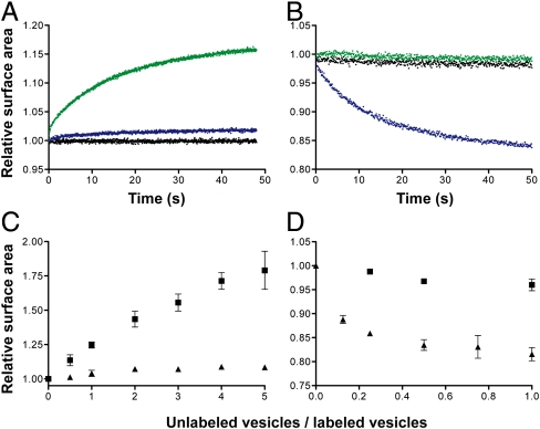
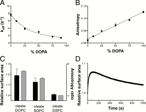
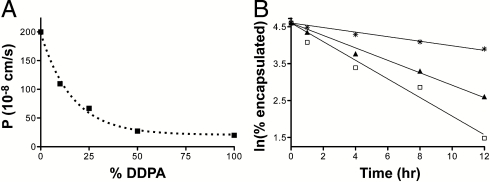
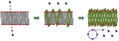
To understand the emergence of Darwinian evolution, it is necessary to identify physical mechanisms that enabled primitive cells to compete with one another. Whereas all modern cell membranes are composed primarily of diacyl or dialkyl glycerol phospholipids, the first cell membranes are thought to have self-assembled from simple, single-chain lipids synthesized in the environment. We asked what selective advantage could have driven the transition from primitive to modern membranes, especially during early stages characterized by low levels of membrane phospholipid. Here we demonstrate that surprisingly low levels of phospholipids can drive protocell membrane growth during competition for single-chain lipids. Growth results from the decreasing fatty acid efflux from membranes with increasing phospholipid content. The ability to synthesize phospholipids from single-chain substrates would have therefore been highly advantageous for early cells competing for a limited supply of lipids. We show that the resulting increase in membrane phospholipid content would have led to a cascade of new selective pressures for the evolution of metabolic and transport machinery to overcome the reduced membrane permeability of diacyl lipid membranes. The evolution of phospholipid membranes could thus have been a deterministic outcome of intrinsic physical processes and a key driving force for early cellular evolution.


Conflict of interest statement

The authors declare no conflict of interest.

Figures

Fig. 1.
Fig. 1.
Phospholipids drive competition between fatty acid vesicles. (A and B) Competition between vesicles was monitored by a FRET-based real-time surface area assay. Growth of FRET-labeled 90∶10 oleate∶DOPA vesicles (A) and shrinkage of FRET-dye labeled oleate vesicles (B) when mixed 1∶1 with buffer (black), unlabeled oleate vesicles (green), or unlabeled 90∶10 oleate∶DOPA vesicles (blue). (C and D) The dependence of vesicle growth or shrinkage on vesicle stoichiometry. Final growth after equilibrium of FRET-labeled 90∶10 oleate∶DOPA vesicles (C) and shrinkage of FRET-labeled oleate vesicles (D) when mixed with varying amounts of unlabeled oleate (▪) or unlabeled 90∶10 oleate∶DOPA (▴) vesicles. Error bars indicate SEM (N = 3).
Fig. 2.
Fig. 2.
Phospholipid-driven growth leads to a filamentous shape transition and vesicle division. (A) Large, multilamellar 90∶10 oleate∶DOPA vesicles, labeled with an encapsulated soluble dye, are initially spherical. (B) Upon mixing with a 100-fold excess of unlabeled oleate vesicles, the mixed vesicles rapidly grow into long, filamentous vesicles. (C) The fragile filamentous vesicles then readily divide into small daughter vesicles upon the application of gentle shear forces (see Methods). (Scale bar: 30 μm.) (D) Time course showing the shape transformation of a labeled 90∶10 oleate∶DOPA vesicle upon addition of unlabeled oleate vesicles. Time in seconds. (Scale bar: 5 μm.)
Fig. 3.
Fig. 3.
Mechanisms of phospholipid-driven growth. (A) The desorption rate of oleate in mixed oleate/DOPA vesicles as a function of DOPA content. Increasing phospholipid content slows oleate desorption, leading to growth of phospholipid-enriched vesicles. (B) The steady-state anisotropy of DPH in oleate/DOPA vesicles as a function of DOPA content. Bilayer packing order increases linearly with increasing fraction of the diacyl lipid. Dashed line indicates linear regression fit, R2 = 0.98. (C) The extent of growth of FRET labeled 90∶10 oleate∶DSPC vesicles when mixed 1∶1 with the given 90∶10 vesicle composition (black bars, left axis) correlates with the ratio of the membrane fluidity between oleate/DSPC bilayers and those of the given composition as measured by DPH anisotropy (gray bars, right axis). DOPC, di-oleoyl-phosphocholine (C18∶1); SOPC, 1-stearoyl-2-oleoyl-phosphocholine (C18∶0/C18∶1). (D) Growth of FRET-labeled 90∶10 oleate∶NA vesicles when mixed 1∶1 with unlabeled oleate vesicles. Growth proceeds in the first 60 s, followed by a slow relaxation due to the equilibration of the slowly exchanging NA fraction. Error bars indicate SEM (N = 3).
Fig. 4.
Fig. 4.
Phospholipids inhibit solute permeation through fatty-acid-based membranes. (A) Permeability of C10 membranes (4∶1∶1 DA∶DOH∶GMD) to ribose as a function of DDPA content as measured by a stopped-flow relaxation assay. (B) Leakage of encapsulated ImpdA from C10 vesicles as measured by scintillation counting of dialysis buffer aliquots. Membrane compositions: □, 4∶1∶1 DA∶DOH∶GMD; ▴, 4∶1∶1 DA∶DOH∶GMD with 25 mol % DDPA; and *, DDPA.
Fig. 5.
Fig. 5.
Schematic for membrane-driven cellular evolution. The gradual transition from highly permeable primitive membranes (Left) to phospholipid membranes (Right) is driven by the selective growth advantage provided by increasing phospholipid content in the membrane. In turn, this transition in membrane composition imposes a selective pressure for the emergence of internalized metabolism to counter the reduced permeability of diacyl lipid membranes.

References

    1. Hargreaves WR, Deamer DW. Liposomes from ionic, single-chain amphiphiles. Biochemistry. 1978;17:3759–3768. - PubMed
    1. Hanczyz MM, Fujikawa SM, Szostak JW. Experimental models of primitive cellular compartmentalization: Encapsulation, growth, and division. Science. 2003;302:618–622. - PMC - PubMed
    1. Mansy SS, et al. Template-directed synthesis of a genetic polymer in a model protocell. Nature. 2008;454:122–125. - PMC - PubMed
    1. Zhu TF, Szostak JW. Coupled growth and division of model protocell membranes. J Am Chem Soc. 2009;131:5705–5713. - PMC - PubMed
    1. Haines TH. Halogen- and sulfur-containing lipids of ochromonas. Annu Rev Microbiol. 1973;27:403–412. - PubMed

Publication types