. 2011 Apr 5;108(14):5813-8.
doi: 10.1073/pnas.1015551108. Epub 2011 Mar 14.

Interaction between hypothalamic dorsomedial nucleus and the suprachiasmatic nucleus determines intensity of food anticipatory behavior

Affiliations

Interaction between hypothalamic dorsomedial nucleus and the suprachiasmatic nucleus determines intensity of food anticipatory behavior

Guadalupe Acosta-Galvan et al. Proc Natl Acad Sci U S A. .

Abstract

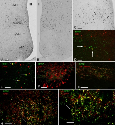
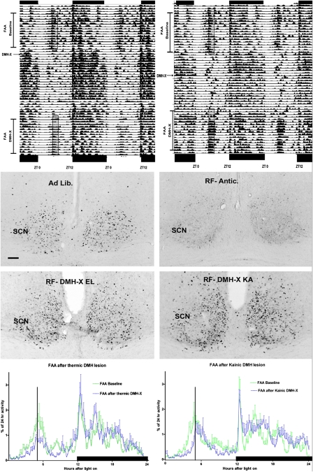
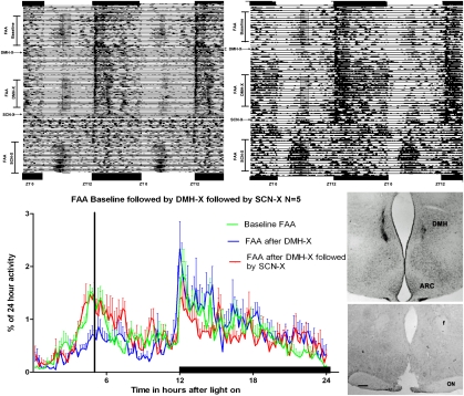
Food anticipatory behavior (FAA) is induced by limiting access to food for a few hours daily. Animals anticipate this scheduled meal event even without the suprachiasmatic nucleus (SCN), the biological clock. Consequently, a food-entrained oscillator has been proposed to be responsible for meal time estimation. Recent studies suggested the dorsomedial hypothalamus (DMH) as the site for this food-entrained oscillator, which has led to considerable controversy in the literature. Herein we demonstrate by means of c-Fos immunohistochemistry that the neuronal activity of the suprachiasmatic nucleus (SCN), which signals the rest phase in nocturnal animals, is reduced when animals anticipate the scheduled food and, simultaneously, neuronal activity within the DMH increases. Using retrograde tracing and confocal analysis, we show that inhibition of SCN neuronal activity is the consequence of activation of GABA-containing neurons in the DMH that project to the SCN. Next, we show that DMH lesions result in a loss or diminution of FAA, simultaneous with increased activity in the SCN. A subsequent lesion of the SCN restored FAA. We conclude that in intact animals, FAA may only occur when the DMH inhibits the activity of the SCN, thus permitting locomotor activity. As a result, FAA originates from a neuronal network comprising an interaction between the DMH and SCN. Moreover, this study shows that the DMH-SCN interaction may serve as an intrahypothalamic system to gate activity instead of rest overriding circadian predetermined temporal patterns.


Conflict of interest statement

The authors declare no conflict of interest.

Figures

Fig. 1.
Fig. 1.
RFRP neurons in the DMH project to the retinorecipient region of the SCN, contain the enzyme for the inhibitory neurotransmitter GABA (GAD), and are activated during food anticipation. (A) Retrograde-labeled neurons in the ventral area of the DMH after an injection of CtB into the SCN. These neurons projecting to the SCN are present in the same ventral area of the DMH that is activated during food anticipation. The distribution of RFRP neurons in the ventral area of the DMH in A coincides with the area expressing c-Fos during FAA in B and coincides with the location of RFRP neurons in the DMH (C). (D) Retrograde-labeled neurons in the DMH after an injection of CtB (red) into the SCN, double-labeled for RFRP (green). (E) Confocal image of RFRP (green) -labeled neurons colocalizing with c-Fos (red) after food restriction. (F) RFRP (green) innervation of the SCN in the area of VIP (red) cellbodies; G shows that this RFRP (green) input is closely associated with retinal fibers (red). H and I illustrate that in the SCN these RFRP (green) fibers colocalize with GAD (red), as is illustrated by the yellow color of green RFRP and red GAD fibers. The arrow indicates the same area in H and I. (Scale bars: A and B, 100 μm; C–F, 50 μm; G and H, 25 μm; I, 10 μm.)
Fig. 2.
Fig. 2.
Diminished food anticipatory behavior after DMH lesion concurs with high neuronal activity in the SCN. (Top)Two double-plotted actograms of locomotor activity illustrate the initial baseline food anticipation (food presented in the time of the translucent rectangle) and the subsequent loss of this anticipatory activity after an electrolytic (Left) or neurotoxic (Right) lesion of the DMH. The activity 2 to 3 h preceding the translucent rectangle is taken as “food anticipatory activity.” (Middle) The neuronal activity by means of c-Fos staining in the SCN 5 h after light onset in an ad libitum control and in a sham restricted-food (RF) animal at the moment of food arrival. The DMH-lesioned animals electrolytic (RF-DMH-X EL) or kainic (RF-DMH-X KA) of the respective actograms above show as high c-Fos expression in the SCN of the ad libitum-fed animal. Only the sham-lesioned food-anticipating animal (RF-antic) showed less c-Fos in the SCN. The quantification of the number of c-Fos–positive neurons in the SCN and the quantification of the FAA for the 2 h before expected food arrival is given in Fig. S5. (Bottom) Waveform analysis of activity during the last 5 d of a restricted-food protocol of the two groups of DMH-X animals illustrating their 24-h activity in 10-min averages. The anticipatory activity to food (as percentage of total 24-h activity) after the lesion is diminished significantly. The vertical bar indicates the time (ZT5) of food delivery (5 h after light onset) and the horizontal black bar the dark period (see Fig. S5 for the quantitative analysis of FAA). (Scale bars, 100 μm.)
Fig. 3.
Fig. 3.
Loss and gain of food anticipation in the same animals bearing, first, a lesion of the DMH, followed by a lesion of the SCN. Both double-plotted actograms depict the activity of animals showing normal anticipatory activity just before the onset of meal delivery (translucent rectangle). This anticipatory activity is nearly completely lost after a kainic acid DMH lesion and returns when the same animal receives an additional SCN lesion. The waveform analysis illustrates the average activity of the same five animals in intact conditions under restricted-food conditions (green line) (the moment of food delivery depicted by the black vertical bar), and illustrates the loss of FAA after DMH-X (blue line) and its return after a successive SCN-X (red line). The right part of the figure shows the injection site of the kainic acid and the section of the hypothalamus showing the lesion of the SCN. The right actogram shows the result of a partial (>70%) lesioned SCN animal that still recovers FAA. See Fig. S9 for the quantification of the activity 3 h before food arrival. (Scale bar, 200 μm.)

References

    1. Schwartz WJ, Reppert SM, Eagan SM, Moore-Ede MC. In vivo metabolic activity of the suprachiasmatic nuclei: A comparative study. Brain Res. 1983;274:184–187. - PubMed
    1. Houben T, Deboer T, van Oosterhout F, Meijer JH. Correlation with behavioral activity and rest implies circadian regulation by SCN neuronal activity levels. J Biol Rhythms. 2009;24:477–487. - PubMed
    1. Stephan FK, Swann JM, Sisk CL. Entrainment of circadian rhythms by feeding schedules in rats with suprachiasmatic lesions. Behav Neural Biol. 1979;25:545–554. - PubMed
    1. Fuller PM, Lu J, Saper CB. Differential rescue of light- and food-entrainable circadian rhythms. Science. 2008;320:1074–1077. - PMC - PubMed
    1. Gooley JJ, Schomer A, Saper CB. The dorsomedial hypothalamic nucleus is critical for the expression of food-entrainable circadian rhythms. Nat Neurosci. 2006;9:398–407. - PubMed

Publication types

MeSH terms