Review
. 2011 Jul;68(7):848-56.
doi: 10.1001/archneurol.2011.46. Epub 2011 Mar 14.

Progenitor cell-based treatment of the pediatric myelin disorders

Affiliations
Review

Progenitor cell-based treatment of the pediatric myelin disorders

Steven A Goldman. Arch Neurol. 2011 Jul.

Abstract

The childhood leukodystrophies are characterized by neonatal or childhood deficiencies in myelin production or maintenance; these may be due to hereditary defects in genes for myelin maintenance, as in Pelizaeus-Merzbacher disease, or to enzymatic deficiencies resulting in substrate misaccumulation or misprocessing, as in the lysosomal storage disorders. Regardless of their respective etiologies, these disorders are essentially all manifested by a profound deterioration in neurological function with age. A congenital deficit in forebrain myelination is also noted in children with the periventricular leukomalacia of cerebral palsy, which yields a more static morbidity. In light of the wide range of disorders to which congenital hypomyelination or postnatal demyelination may contribute, and the relative homogeneity of oligodendrocytes and their progenitors, the leukodystrophies may be especially attractive targets for cell-based therapeutic strategies. As a result, glial progenitor cells, which can give rise to new myelinogenic oligodendrocytes, have become of great interest as potential vectors for the restoration of myelin to the dysmyelinated brain and spinal cord. In addition, by distributing throughout the neuraxis after perinatal graft, and giving rise to astrocytes as well as oligodendrocytes, glial progenitor cells may be of great utility in rectifying the dysmyelination-associated enzymatic deficiencies of the lysosomal storage disorders.


Figures
Figure
Figure
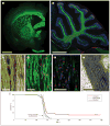
Myelination of a leukodystrophic brain by engrafted human glial progenitor cells. Implanted human fetal glial progenitor cells myelinated extensive regions of a shiverer mouse forebrain. These images are taken from either 1-year-old (A and B), 12-week-old (C and D), or 35-week-old (E and F) immunodeficient and myelin-deficient shiverer mice (shi/shi × rag2−/−), implanted at birth with A2B5+/PSA-NCAM-–sorted human glial progenitor cells. A, Low-magnification coronal image of the shiverer forebrain after transplant, immunostained for myelin basic protein (MBP) (green); because these mice are MBP deficient, all observed MBP immunoreactivity is of human origin. B, Higher-magnification view of the engrafted shiverer cerebellum, illustrating the high-efficiency myelination of the cerebellar white matter (MBP, green) by human glial progenitor cells (red) (4′,6-diamidino-2-phenylindole counterstain of mouse cells, blue). C, Individual myelinated human oligodendrocytes (human nuclear antigen, red; MBP, green) in the callosum of a 12-week-old shiverer who underwent neonatal transplant. D, Confocal image showing the donor myelin (MBP, red)–ensheathed host axons (neurofilament, green) imaged in the cervical spinal cord of a 1-year-old shiverer × rag2–null mouse who underwent transplant. E, Optical section through the cerebellar white matter of the 35-week-old shiverer who underwent transplant, manifesting the normal nodal organization of its donor-myelinated axons. Caspr (a paranodal protein), red; Caspr2 (a juxtaparanodal marker), green. F, Electron micrograph of a myelinated fiber in the callosum of a shiverer brain, 12 weeks after neonatal graft, showing the normal major dense lines and myelin lamellae of the donor cell–myelinated recipient axon. G, Kaplan-Meier plot showing the survival curves of shiverer mice who were untreated, were treated with a saline control, or underwent human glial progenitor cell transplant. Whereas all untreated shiverer mice invariably died by 21 weeks of age, a little less than a fourth of mice who received neonatal xenograft lived, some for as long as 2 years after birth, with substantial recovery of lost neurological function. Adapted from Windrem et al., Scale bar=1 mm in parts A and B; 10 μm in parts C and D; 5 μm in part E; and 1 μm in part F.

References

    1. Goldman SA. Stem and progenitor cell-based therapy of the human central nervous system. Nat Biotechnol. 2005;23(7):862–871. - PubMed
    1. Goldman SA, Windrem MS. Cell replacement therapy in neurological disease. Philos Trans R Soc Lond B Biol Sci. 2006;361(1473):1463–1475. - PMC - PubMed
    1. Koch P, Kokaia Z, Lindvall O, Brüstle O. Emerging concepts in neural stem cell research: autologous repair and cell-based disease modelling. Lancet Neurol. 2009;8(9):819–829. - PubMed
    1. Lindvall O, Kokaia Z. Stem cells in human neurodegenerative disorders: time for clinical translation? J Clin Invest. 2010;120(1):29–40. - PMC - PubMed
    1. Martino G, Franklin RJ, Van Evercooren AB, Kerr DA Stem Cells in Multiple Sclerosis (STEMS) Consensus Group. Stem cell transplantation in multiple sclerosis: current status and future prospects. Nat Rev Neurol. 2010;6(5):247–255. - PubMed

Publication types