. 2011 Mar 29;123(12):1287-96.
doi: 10.1161/CIRCULATIONAHA.110.982918. Epub 2011 Mar 14.

Human cardiac stem cell differentiation is regulated by a mircrine mechanism

Affiliations

Human cardiac stem cell differentiation is regulated by a mircrine mechanism

Toru Hosoda et al. Circulation. .

Erratum in

  • Correction.
    [No authors listed] [No authors listed] Circulation. 2015 Dec 1;132(22):e362. doi: 10.1161/CIR.0000000000000340. Circulation. 2015. PMID: 26621665 No abstract available.

Retraction in

Expression of concern in

  • Expression of Concern.
    [No authors listed] [No authors listed] Circ Res. 2019 Jan 18;124(2):e4-e5. doi: 10.1161/RES.0000000000000241. Circ Res. 2019. PMID: 30582460 No abstract available.
  • Expression of Concern.
    [No authors listed] [No authors listed] Circulation. 2019 Jan 15;139(3):e5-e6. doi: 10.1161/CIR.0000000000000639. Circulation. 2019. PMID: 30615475 No abstract available.

Abstract

Background: Cardiac stem cells (CSCs) delivered to the infarcted heart generate a large number of small fetal-neonatal cardiomyocytes that fail to acquire the differentiated phenotype. However, the interaction of CSCs with postmitotic myocytes results in the formation of cells with adult characteristics.

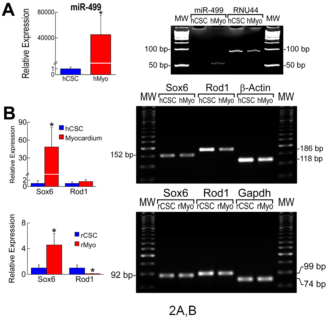
Methods and results: On the basis of results of in vitro and in vivo assays, we report that the commitment of human CSCs (hCSCs) to the myocyte lineage and the generation of mature working cardiomyocytes are influenced by microRNA-499 (miR-499), which is barely detectable in hCSCs but is highly expressed in postmitotic human cardiomyocytes. miR-499 traverses gap junction channels and translocates to structurally coupled hCSCs favoring their differentiation into functionally competent cells. Expression of miR-499 in hCSCs represses the miR-499 target genes Sox6 and Rod1, enhancing cardiomyogenesis in vitro and after infarction in vivo. Although cardiac repair was detected in all cell-treated infarcted hearts, the aggregate volume of the regenerated myocyte mass and myocyte cell volume were greater in animals injected with hCSCs overexpressing miR-499. Treatment with hCSCs resulted in an improvement in ventricular function, consisting of a better preservation of developed pressure and positive and negative dP/dt after infarction. An additional positive effect on cardiac performance occurred with miR-499, pointing to enhanced myocyte differentiation/hypertrophy as the mechanism by which miR-499 potentiated the restoration of myocardial mass and function in the infarcted heart.

Conclusions: The recognition that miR-499 promotes the differentiation of hCSCs into mechanically integrated cardiomyocytes has important clinical implications for the treatment of human heart failure.


Figures

Figure 1
Figure 1
Expression of miRs in cardiomyocytes and CSCs. A, Expression of selected miRs in rat cardiomyocytes (Myo) and CSCs. B and C, miR-499 in rCSCs and rMyo parallels Myh7b mRNA expression. Small nucleolar snoRNA, normalization. Gapdh: housekeeping-gene.
Figure 2
Figure 2
Target genes of miR-499. A, miR-499 in hCSCs and human-cardiomyocytes (hMyo). Small nucleolar RNA RNU44, normalization. B, Sox6 and Rod1 mRNA. Fold-changes vs. hCSCs or rCSCs. C, Protein levels of Sox6 and Rod1. Loading, Gapdh and Ponceau red. D, qRT-PCR of miR-499 in transfected 3T3 cells. E, Repressive activity of miR-499 on Sox6 and Rod1 3’-UTRs by luciferase assay. *,**,†P<0.05 vs. 0, 0.1 and 0.4 µg, respectively.
Figure 2
Figure 2
Target genes of miR-499. A, miR-499 in hCSCs and human-cardiomyocytes (hMyo). Small nucleolar RNA RNU44, normalization. B, Sox6 and Rod1 mRNA. Fold-changes vs. hCSCs or rCSCs. C, Protein levels of Sox6 and Rod1. Loading, Gapdh and Ponceau red. D, qRT-PCR of miR-499 in transfected 3T3 cells. E, Repressive activity of miR-499 on Sox6 and Rod1 3’-UTRs by luciferase assay. *,**,†P<0.05 vs. 0, 0.1 and 0.4 µg, respectively.
Figure 3
Figure 3
Translocation of miR-499 through gap junctions. A, qRT-PCR of miR-499 in transfected C2C12 cells (+miR). C2C12: non-transfected cells. B, Co-culture of C2C12 cells and hCSCs. Upper panels: hCSCs show red and green fluorescence (miR-499-EGFP, arrows) and C2C12 cells show red fluorescence only (miR-499). Lower panels: connexin 43 (white, arrowheads) at the interface of hCSCs and C2C12 cells. C, Fraction of hCSCs expressing miR-499. +α-GA, gap junction inhibitor. *P<0.05 vs. -α-GA. D, miR-499 expression in hCSCs seeded alone (Control), hCSCs exposed to medium collected from myocyte cultures (+Medium), EGFP-positive hCSCs sorted by FACS 5 days after co-culture with myocytes in the absence (+Myo) and presence (+Myo +α-GA) of α-GA. *,**,†P<0.05 vs. Control, +Medium, and +Myo, respectively. E, Co-culture of cardiomyocytes (α-SA, yellow) and EGFP-positive hCSCs (green). At 2 hours (left panel), miR-499 (red) is confined to myocytes. At 36 hours (central panel), hCSCs show miR-499 (red) and EGFP (miR-499-EGFP, arrowheads). Connexin 43 is present at the interface of hCSCs and myocytes (white). Scrambled probe (right panel). F, Results are mean±SD. +α-GA, gap junction inhibitor, 10 µM. *P<0.05 vs. -α-GA. G, hCSC and myocyte apoptosis and α-GA. *,**P<0.05 vs. Control and 10 µM α-GA, respectively. H, Repressive activity of translocated miR-499 on Sox6 UTR in hCSCs is abolished by α-GA, 10 µM. Blank: donor cells transfected with empty plasmid. *,**P<0.05 vs. blank and miR-499, respectively.
Figure 3
Figure 3
Translocation of miR-499 through gap junctions. A, qRT-PCR of miR-499 in transfected C2C12 cells (+miR). C2C12: non-transfected cells. B, Co-culture of C2C12 cells and hCSCs. Upper panels: hCSCs show red and green fluorescence (miR-499-EGFP, arrows) and C2C12 cells show red fluorescence only (miR-499). Lower panels: connexin 43 (white, arrowheads) at the interface of hCSCs and C2C12 cells. C, Fraction of hCSCs expressing miR-499. +α-GA, gap junction inhibitor. *P<0.05 vs. -α-GA. D, miR-499 expression in hCSCs seeded alone (Control), hCSCs exposed to medium collected from myocyte cultures (+Medium), EGFP-positive hCSCs sorted by FACS 5 days after co-culture with myocytes in the absence (+Myo) and presence (+Myo +α-GA) of α-GA. *,**,†P<0.05 vs. Control, +Medium, and +Myo, respectively. E, Co-culture of cardiomyocytes (α-SA, yellow) and EGFP-positive hCSCs (green). At 2 hours (left panel), miR-499 (red) is confined to myocytes. At 36 hours (central panel), hCSCs show miR-499 (red) and EGFP (miR-499-EGFP, arrowheads). Connexin 43 is present at the interface of hCSCs and myocytes (white). Scrambled probe (right panel). F, Results are mean±SD. +α-GA, gap junction inhibitor, 10 µM. *P<0.05 vs. -α-GA. G, hCSC and myocyte apoptosis and α-GA. *,**P<0.05 vs. Control and 10 µM α-GA, respectively. H, Repressive activity of translocated miR-499 on Sox6 UTR in hCSCs is abolished by α-GA, 10 µM. Blank: donor cells transfected with empty plasmid. *,**P<0.05 vs. blank and miR-499, respectively.
Figure 4
Figure 4
Function of miR-499 in hCSCs. A, Sox6 and Rod1 expression in hCSCs infected with a lentivirus carrying DsRed only (control) or DsRed-miR-499. Loading: Ponceau red. *P<0.05 vs. Control. B, hCSCs positive for BrdU, Nkx2.5 and GATA4. *P < 0.05 vs. Control. C, Sox6 and Rod1 mRNA in hCSCs transfected with negative siRNA (Neg) or specific siRNA (Spec). *P<0.05 vs. Neg. D, hCSCs positive for Nkx2.5 and GATA4. *P<0.05 vs. siRNA-Neg.
Figure 5
Figure 5
Cardiac repair with miR-499-EGFP-hCSCs. A, Regenerated myocardium (arrowheads) within the infarct 10 days after surgery and cell implantation. The area included in the rectangle is shown at higher magnification in the inset. Myocytes: α-sarcomeric-actin, α-SA, red. EGFP: green. *Dead myocytes. B and C, Localization of BrdU (B, red) and Ki67 (C, magenta) in EGFP-positive human-myocytes. Cell boundaries are defined by laminin (white). D, Human-myocytes, stained by α-SA (red) and EGFP (green), exhibit sarcomere striation and are positive for Alu (white).
Figure 6
Figure 6
Regenerated myocytes. A, Connexin 43 and N-cadherin (white) are expressed in EGFP-positive α-SA-positive human-myocytes (arrowheads). B, Aggregate myocyte volume and average cell volume of human-myocytes. Infarcted hearts treated with EGFP-hCSCs (Ctrl) or miR-499-EGFP-hCSCs (miR). *P<0.05 vs. Ctrl. C, Size distribution of human-myocytes derived from differentiation of EGFP-hCSCs (green-bars) and miR-499-EGFP-hCSCs (red-bars).
Figure 7
Figure 7
Ventricular function and mechanics of human myocytes. A, LV end-diastolic pressure (LVEDP), LV systolic pressure (LVSP), LV developed pressure (LVDevP), and LV+ and -dP/dt in sham-operated (Sham), infarcted-untreated (MI), and infarcted hearts treated with EGFP-hCSCs (Ctrl) or miR-499-EGFP-hCSCs (miR). *,**,†P<0.05 vs. Sham, MI and Ctrl, respectively. B, Human-myocytes derived from the differentiation of EGFP-hCSCs (left panels) and miR-499-EGFP-hCSCs (right panels). C, Volume of myocytes in Ctrl and miR. D, Fractional-shortening of regenerated human-myocytes (New) and surviving rat-myocytes (Surv): tracings of surviving and regenerated myocytes derived from miR-499-EGFP-hCSCs. Spared myocytes had depressed fractional shortening. *P<0.05 vs. Surv.

Comment in

References

    1. Hosoda T, D’Amario D, Zheng H, Padin-Iruegas ME, Yasuzawa S, Amano K, Cheng W, Urbanek K, Rota M, Kajstura J, Anversa P, Leri A. Clonality of mouse and human cardiomyogenesis in vivo. Proc Natl Acad Sci USA. 2009;106:17169–17174. - PMC - PubMed
    1. Gonzalez A, Rota M, Nurzynska D, Misao Y, Tillmanns J, Ojaimi C, Padin-Iruegas ME, Müller P, Esposito G, Bearzi C, Vitale S, Dawn B, Sanganalmath SK, Baker M, Hintze TH, Bolli R, Urbanek K, Hosoda T, Anversa P, Kajstura J, Leri A. Activation of cardiac progenitor cells reverses the failing heart senescent phenotype and prolongs lifespan. Circ Res. 2008;102:597–606. - PubMed
    1. Pfister O, Mouquet F, Jain M, Summer R, Helmes M, Fine A, Colucci WS, Liao R. CD31− but Not CD31+ cardiac side population cells exhibit functional cardiomyogenic differentiation. Circ Res. 2005;97:52–61. - PubMed
    1. Bearzi C, Rota M, Hosoda T, Tillmanns J, Nascimbene A, De Angelis A, Yasuzawa-Amano S, Trofimova I, Siggins RW, Lecapitaine N, Cascapera S, Beltrami AP, D'Alessandro DA, Zias E, Quaini F, Urbanek K, Michler RE, Bolli R, Kajstura J, Leri A, Anversa P. Human cardiac stem cells. Proc Natl Acad Sci USA. 2007;104:14068–14073. - PMC - PubMed
    1. Boni A, Urbanek K, Nascimbene A, Hosoda T, Zheng H, Delucchi F, Amano K, Gonzalez A, Vitale S, Ojaimi C, Rizzi R, Bolli R, Yutzey KE, Rota M, Kajstura J, Anversa P, Leri A. Notch1 regulates the fate of cardiac progenitor cells. Proc Natl Acad Sci USA. 2008;105:15529–15534. - PMC - PubMed

Publication types

MeSH terms