. 2011;25(2):279-93.
doi: 10.3233/JAD-2011-101014.

Microglia demonstrate age-dependent interaction with amyloid-β fibrils

Affiliations

Microglia demonstrate age-dependent interaction with amyloid-β fibrils

Angela Marie Floden et al. J Alzheimers Dis. 2011.

Abstract

Alzheimer's disease (AD) is an age-associated disease characterized by increased accumulation of extracellular amyloid-β (Aβ) plaques within the brain. Histological examination has also revealed profound microglial activation in diseased brains often in association with these fibrillar peptide aggregates. The paradoxical presence of increased, reactive microglia yet accumulating extracellular debris suggests that these cells may be phagocytically compromised during disease. Prior work has demonstrated that primary microglia from adult mice are unable to phagocytose fibrillar Aβ1-42 in vitro when compared to microglia cultured from early postnatal animals. These data suggest that microglia undergo an age-associated decrease in microglial ability to interact with Aβ fibrils. In order to better define a temporal profile of microglia-Aβ interaction, acutely isolated, rather than cultured, microglia from 2 month, 6 month, and postnatal day 0 C57BL/6 mice were compared. Postnatal day 0 microglia demonstrated a CD47 dependent ability to phagocytose Aβ fibrils that was lost by 6 months. This corresponded with the ability of postnatal day 0 but not adult microglia to decrease Aβ immunoreactive plaque load from AD sections in vitro. In spite of limited Aβ uptake ability, adult microglia had functional phagocytic uptake of bacterial bioparticles and demonstrated the ability to adhere to both Aβ plaques and in vitro fibrillized Aβ. These data demonstrate a temporal profile of specifically Aβ-microglia interaction with a critical developmental period at 6 months in which cells remain able to interact with Aβ fibrils but lose their ability to phagocytose it.


Figures
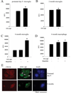
Figure 1
Figure 1
Microglia demonstrated decreased ability to phagocytose Aβ fibrils with age. Acutely isolated microglia from (A) postnatal day 0, (B) 2 month, (C) 6 month, or (D) peritoneal macrophage from 6 month mice were incubated with 500nM FITC-Aβ or 0.125mg/mL FITC-bioparticles (positive control) for 6 hours. Intracellular relative fluorescence units (RFU) were quantitated and averaged via plate reader (480nm excitation and 520nm emission) and averaged (+/−SD). Graphs are representative of 4 independent experiments analyzed by t-test or one-way ANOVA (*p<0.001 from control). (E) Postnatal day 0 microglia were incubated with 500nM FITC-Aβ for 6 hours then fixed and immunostained using anti-CD11b antibody and texas red-conjugated secondary antibody with DAPI counterstain for nuclei. Intracellular Aβ (arrows) were visualized via FITC conjugation.
Figure 2
Figure 2
Selected Aβ receptor protein levels were compared between cultured and acutely isolated mouse microglia with age. Microglia were isolated from mixed cultures at 14 days in vitro (cultured) and stimulated 24hr with or without 25ng/mL LPS (cultured+LPS) or acutely from postnatal day 0 (P0) 2–4 month, 6–8 month, or 12–17 month old mice and Western blotted for quantitation of optical densities (O.D.) of selected proteins (α6 integrin, β1 integrin, TLR2, CD36, CD47, SRA-1, FPRL1, LRP, and RAGE) normalized against their respective loading controls (ERK2). Microglia were isolated from 10 animals per age group and pooled for each experiment. Experiments were repeated 3–5 times (totaling 30–50 animals per condition) and averaged +/−SD. Statistical significance was determined via one-way ANOVA (*p<0.05 from P0).
Figure 3
Figure 3
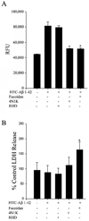
Acutely isolated microglia from postnatal day 0 mice take up Aβ fibrils in a CD47 but not a scavenger receptor or β1 integrin-dependent manner. Postnatal day 0 (P0) acutely isolated microglia were incubated with specific antagonists for scavenger-type receptors (300µg/mL Fucoidan), CD47 (100µg/mL 4N1K), or β1 integrin (100µg/mL RHD-containing peptide). Cells were incubated with and without antagonists for 30 minutes, 37°C, then incubated with 1µM FITC-Aβ 1–42 for 2 hours. (A) After incubation, relative intracellular fluorescence units (RFU) were read (480nm excitation and 520nm emission) via plate reader. (B) To determine cell viability after the 2 hour stimulation, cellular release of lactate dehydrogenase (LDH) was measured from culture media. Graphs are average values (±SD) and representative of 5 independent experiments analyzed via one-way ANOVA (*p<0.001 from Aβ control).
Figure 4
Figure 4
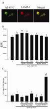
A CD47 receptor antagonist dose-dependently attenuated the ability of acutely isolated postnatal day 0 microglia to take up Aβ fibrils. Microglia from P0 C57BL/6 pups were acutely isolated from brains and cultured for 6 hours with 1µM FITC-Aβ. (A) Cells were fixed and immunostained using an anti-LAMP-1 antibody. Antibody binding was visualized using a texas red conjugated secondary antibody. (B,C) Alternatively, microglia were pretreated with 10, 100, or 500µg/mL Fucoidan or 4N1K for 30 min. prior to the 6 hour 1µM Aβ-FITC stimulation. (B) After the 6 hour stimulation, relative fluorescence intensity units (RFU) from phagocytosed peptide in each condition were quantitated via plate reader (480 nm excitation and 520nm emission) and averaged (± SD). (C) Cell viability after the 6 hour stimulation was determined by quantitating LDH released into the media normalized to cellular LDH levels and graphed as percent control release. Graphs are representative of three independent experiments analyzed via one-way ANOVA. (%p<0.01; *p<0.05; **p<0.001 from control; $p<0.01; &p<0.05; #p<0.001 from Aβ).
Figure 5
Figure 5
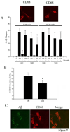
Postnatal day 0 but not 6 month old microglia decreased numbers of Aβ plaques when co-cultured with AD brain tissue. (A) Human temporal cortex from age-matched control or AD tissue (n=3) was cultured with or without acutely isolated microglia (35,000 cells/well) from P0 or 6 month C57BL/6 mice for 3 or 7 days in vitro (DIV). The cultures were fixed at each time point and immunostained with anti-mouse CD68 antibody to visualize exogenous murine microglia as well as with anti-Aβ antibody to detect Aβ containing plaques. Plaque numbers were counted, averaged ±SD, analyzed via one-way ANOVA, and graphed. (*p<0.001 from 7 day no microglia AD tissue only). (B) To compare microglia viability after 7 DIV, microglia from acutely isolated postnatal day 0 and 6 month old mice were cultured without tissue for 7 days. Cells were fixed, immunostained using an anti-CD68 antibody, and counted and averaged ±SD (*p<0.005 from P0). Graphs are representative of 3 independent experiments analyzed via t-test. (C) Representative images are shown of postnatal day 0 microglia cultured on an AD brain section for 7 days in vitro double-stained with anti-mouse CD68 and texas red-conjugated secondary antibody along with anti-Aβ antibody and FITC-conjugated secondary antibody to visualize Aβ uptake into the cells.
Figure 6
Figure 6
Microglia isolated from 6 month and postnatal day 0 microglia were adherent to Aβ plaques and Aβ fibrils in vitro. Microglia from P0 or 6–8 month old C57BL/6 mice were acutely isolated from brains, labeled with 3µM DAPI (blue nuclei), and seeded onto unfixed AD or age-matched control cryosections (10µm) for 1hr. The tissue was rinsed to remove unbound cells and and the remaining cells and tissue were fixed and plaques immunostained using an anti-Aβ antibody. (A) A representative image of AD plaque-adherent P0 and 6 month microglia is shown (arrows). Scale bar, 100µm. (B) The number of microglia adherent to Aβ immunoreactive plaques were averaged ± SD, analyzed via one-way ANOVA and graphed. Data is representative of 4 independent experiments. (C) Alternatively, 0.125nM/mm2 fibrillized Aβ was coated onto a 96 well plate and dried. Microglia from postnatal day 0 or 6 month animals were isolated, prelabeled with 3µm DAPI, and allowed to adhere to tissue culture plastic only (Control) or Aβ coated wells for 1 hour. The wells were rinsed to clear any unbound microglia and the fluorescence intensity was quantitated via plate reader (346 nm excitation and 460 nm emission), averaged ± SD and analyzed by one-way ANOVA. The data shown is representative of 3 independent experiments.

References

    1. McGeer PL, Itagaki S, Tago H, McGeer EG. Reactive microglia in patients with senile dementia of the Alzheimer type are positive for the histocompatibility glycoprotein HLA-DR. Neurosci Lett. 1987;79:195–200. - PubMed
    1. Styren SD, Civin WH, Rogers J. Molecular, cellular, and pathologic characterization of HLA-DR immunoreactivity in normal elderly and Alzheimer's disease brain. Exp Neurol. 1990;110:93–104. - PubMed
    1. Akiyama H, McGeer PL. Brain microglia constitutively express beta-2 integrins. J Neuroimmunol. 1990;30:81–93. - PubMed
    1. Cras P, Kawai M, Siedlak S, Mulvihill P, Gambetti P, Lowery D, Gonzalez-DeWhitt P, Greenberg B, Perry G. Neuronal and microglial involvement in beta-amyloid protein deposition in Alzheimer's disease. Am J Pathol. 1990;137:241–246. - PMC - PubMed
    1. Sasaki A, Yamaguchi H, Ogawa A, Sugihara S, Nakazato Y. Microglial activation in early stages of amyloid beta protein deposition. Acta Neuropathol. 1997;94:316–322. - PubMed

Publication types

MeSH terms