Comparative Study
. 2011 May 15;91(9):968-75.
doi: 10.1097/TP.0b013e318212c7fe.

Comparison of Gal and non-Gal-mediated cardiac xenograft rejection

Affiliations
Comparative Study

Comparison of Gal and non-Gal-mediated cardiac xenograft rejection

Henry D Tazelaar et al. Transplantation. .

Abstract

Background: This study compares the pathologic condition of delayed xenograft rejection in Gal-positive and Gal-knockout cardiac xenografts after pig-to-baboon heterotopic cardiac xenotransplantation when the induced anti-Gal antibody response is unregulated, blocked, or absent.

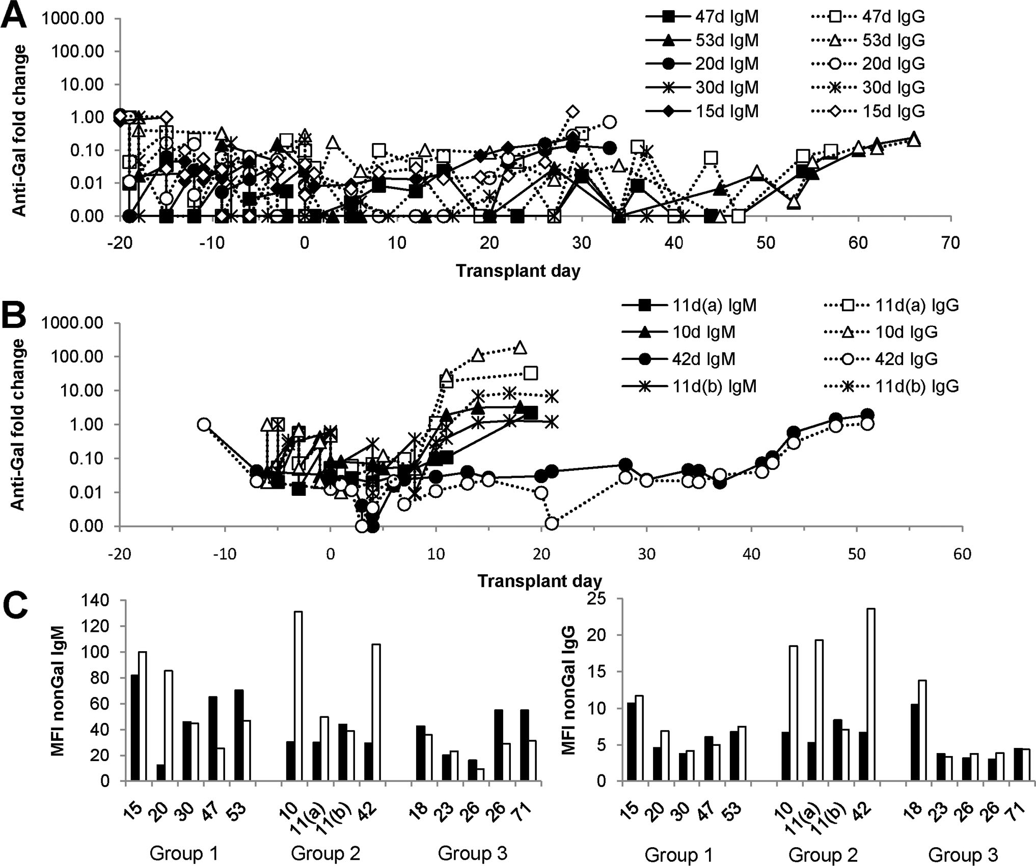
Methods: Baboon recipients of Gal-positive, CD46 pig hearts were treated with an αGal polymer (group 1; n=11) or Gal-specific immunoapheresis (group 2; n=8) to block anti-Gal antibody. Gal-knockout cardiac xenografts recipients (group 3; n=5) received no anti-Gal therapy. Perioperative and interim biopsies were examined and antibody responses were determined.

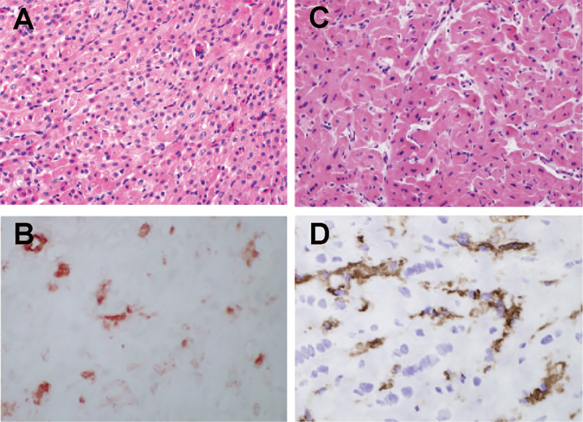
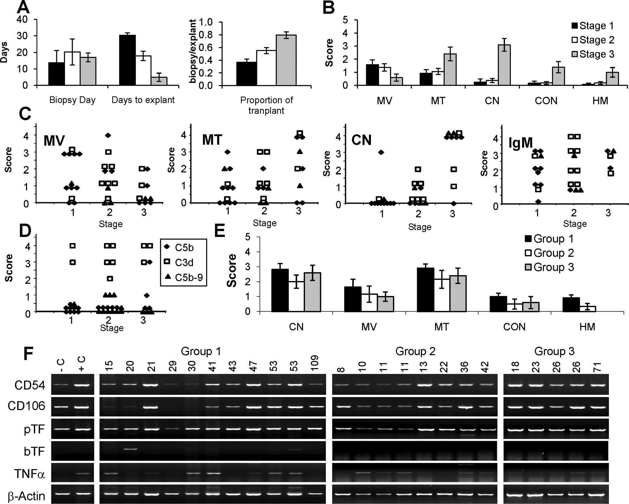
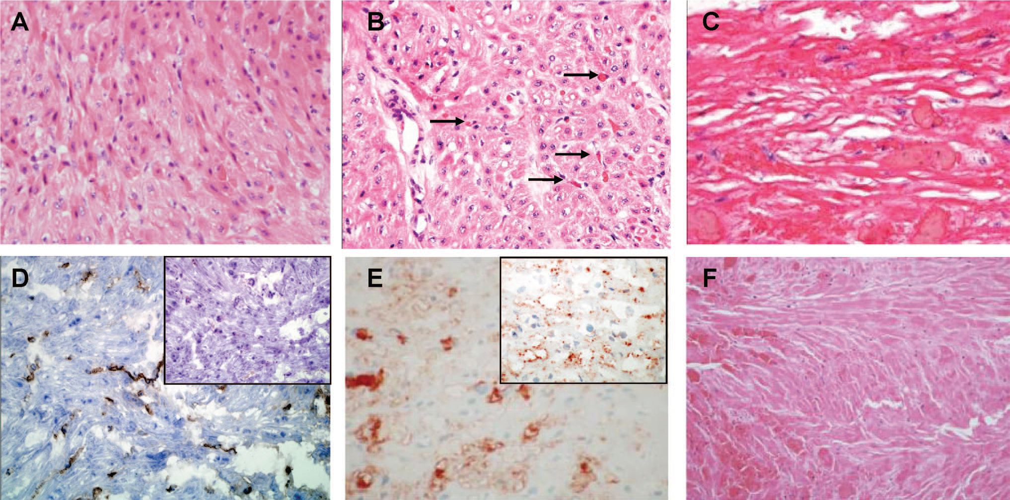
Results: No hyperacute rejection was seen and histologic findings were similar across the groups. All groups showed vascular antibody deposition in perioperative and interim biopsies and in explant samples. A prominent antibody response was detected only in group 2. Complement activation was evident by C3d deposition but deposition of C5b and C5b-9 was limited. Earliest evidence of myocardial injury was myocyte vacuolization in the absence of microvascular thrombosis or coagulative necrosis that developed later. Histology of explanted hearts exhibited mainly microvascular thrombosis and coagulative necrosis with little evidence of interstitial hemorrhage or edema.

Conclusions: The histology of rejection seemed independent of the anti-Gal or non-Gal immune response. Myocyte vacuolization seems to be an early feature of delayed xenograft rejection presaging more classic pathologic features.


Figures

FIGURE 1.
FIGURE 1.
Antibody response. The graphs depict the variation in anti-Gal (A and B) and non-Gal (C) antibody after cardiac xenotransplantation. Anti-Gal IgM and IgG were measured by enzyme-linked immunosorbent assay as previously described (38) and is depicted as the fold change compared with pretransplant serum. Non-Gal antibody was determined using flow cytometry as indicated in the Materials and Methods. (A) Group 1 anti-Gal IgM and IgG. (B) Group 2 anti-Gal IgM and IgG. A and B show antibody responses in all recipients who survived xenograft explant. The day of graft rejection for each recipient is indicated in the figure. (C) Non-Gal immune responses in groups 1–3. The graph compares non-Gal IgM (left) and IgG (right) levels in pretransplant serum (filled bars) with postexplant or necropsy serum (open bars). The numbers below the bar graphs indicate the duration of the transplant for each study group (groups 1, 2, and 3) and correspond to the values in A and B for groups 1 and 2. No group 3 recipient survived explant, so the data compare non-Gal antibody levels in pretransplant and necropsy serum.
FIGURE 2.
FIGURE 2.
Xenograft histology in 30-min biopsies. (A, B) GT+ cardiac xenograft biopsy; (C, D) GTKO biopsy; (A, C) hematoxylin-eosin stain; (B, D) IgM staining. The IgM staining in B and D was scored diffuse 2. Parallel staining for C5d was negative (data not shown).
FIGURE 3.
FIGURE 3.
Staging and histologic features of interim biopsies and explanted left ventricle cross-sections. (A) Biopsies were staged based on levels of cardiac contraction and serum troponin (22). Stages 1 (black filled bars), 2 (white filled bars), and 3 (gray filled bars) were independent of the average biopsy day but were inversely related to the days to explants (explants day–biopsy day) and directly related to the proportion of transplant (biopsy day/explants day). (B) Average stage-dependent histologic features of interim biopsies. Each bar is the mean histologic score. (C) Stage-specific changes in interim biopsy histology for transplant group 1 (filled diamond), 2 (open square), and 3 (filled triangle). The heading for each panel (MV, MT, and CN) is the same as in (B). The IgM heading denotes vascular IgM staining. (D) Stage-specific vascular deposition of C3d, C5b, and C5b-9. (E) Average histology scores in each transplant group for explanted left ventricle cross-sections. Abbreviations are the same as in B and F. Semiquantitative reverse-transcriptase polymerase chain reaction of intragraft expression for porcine intracellular adhesion molecule CD54, vascular cellular adhesion molecule CD106, porcine tissue factor (pTF) and baboon tissue factor (bTF), and tumor necrosis factor (TNF)-α. Negative control (C) is a normal pig heart. Positive control sample (C) is an explanted pig heart after pig-to-primate heterotopic xenotransplantation performed without immunosuppression. The transplant group and survival times are indicated above each lane. In A, B, and E, the error bars are standard error of the mean. MV, myocyte vacuolization; MT, microvascular thrombosis; CN, coagulative necrosis; CON, vascular congestion; HM, hemorrhage.
FIGURE 4.
FIGURE 4.
Histology of interim biopsies and explanted tissue. (A) Hematoxylin-eosin (H&E) staining of stage 1 biopsy from a group 1 recipient. High levels of myocyte vacuolization are present with minimal microvascular thrombosis or coagulative necrosis. (B) H&E stain of stage 2 biopsy from a group 1 transplant showing continued myocyte vacuolization with increased microvascular thrombosis (arrows) and low levels of coagulative necrosis. (C) H&E of stage 3 biopsy from a group 1 transplant showing widespread coagulative necrosis. (D) Vascular IgM deposition in a stage 1 biopsy from group 3. The inset shows negative staining for C5b. (E) Vascular IgM staining in a stage 2 biopsy from group 2. The inset shows positive C3d staining. (E) H&E staining of a group 1 explanted left ventricle rejected on postoperative day 53.

References

    1. Platt JL, Lin SS, McGregor CGA. Acute vascular rejection. Xenotransplantation 1998; 5: 169. - PubMed
    1. Platt JL, Fischel RJ, Matas AJ, et al. Immunopathology of hyperacute xenograft rejection in a swine-to-primatemodel. Transplantation 1991; 52: 214. - PubMed
    1. Schuurman H-J, Cheng J, Lam T. Pathology of xenograft rejection: A commentary. Xenotransplantation 2003; 10: 293. - PubMed
    1. Weisman HF, Bartow T, Leppo MK, et al. Soluble human complement receptor type 1: In vivo inhibitor of complement suppressing post-ischemic myocardial inflammation and necrosis. Science 1990; 249: 146. - PubMed
    1. Miyagawa S, Hirose H, Shirakura R, et al. The mechanism of discordant xenograft rejection. Transplantation 1988; 46: 825. - PubMed

Publication types

MeSH terms

Substances