. 2011:2011:405437.
doi: 10.1155/2011/405437. Epub 2011 Feb 15.

Chondrosarcoma: with updates on molecular genetics

Affiliations

Chondrosarcoma: with updates on molecular genetics

Mi-Jung Kim et al. Sarcoma. 2011.

Abstract

Chondrosarcoma (CHS) is a malignant cartilage-forming tumor and usually occurs within the medullary canal of long bones and pelvic bones. Based on the morphologic feature alone, a correct diangosis of CHS may be difficult, Therefore, correlation of radiological and clinicopathological features is mandatory in the diagnosis of CHS. The prognosis of CHS is closely related to histologic grading, however, histologic grading may be subjective with high inter-observer variability. In this paper, we present histologic grading system and clinicopathological and radiological findings of conventional CHS. Subtypes of CHSs, such as dedifferentiated, mesenchymal, and clear cell CHSs are also presented. In addition, we introduce updated cytogenetic and molecular genetic findings to expand our understanding of CHS biology. New markers of cell differentiation, proliferation, and cell signaling might offer important therapeutic and prognostic information in near future.


Figures
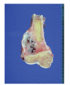
Figure 1
Figure 1
A well-demarcated cartilaginous tumor, measuring 6.5 cm in greatest dimension, in the metaphysis of distal femur. The mass extends through the periosteum to adjacent soft tissue.
Figure 2
Figure 2
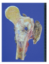
An intramedullary cartilaginous mass, measuring 9.5 cm in greatest dimension, in the metadiaphysis of proximal femur. Note the white to bluish gray and lobulated cut surface and cortical destruction.
Figure 3
Figure 3
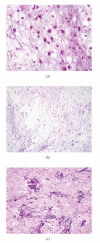
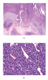
(a) Atypical chondrocytes with binucleation and eosinophilic cytoplasm; (b) chondrosarcoma with myxoid change; (c) chondrosarcoma with necrotic tumor cells and calcification.
Figure 4
Figure 4
(a) Grade 1 CHS with chondroid matrix and low cellularity. Note the soft tissue extension. (b) Grade 2 CHS with increased cellularity and soft tissue extension. (c) Grade 3 CHS with more increased cellularity and cellular atypia. (d) Pleomorphic tumor cells and frequent multinucleated cells in grade 3 CHS.
Figure 5
Figure 5
Schematic representation illustrating grading system of conventional CHS (not all CHSs follow this scheme).
Figure 6
Figure 6
CHS permeating between preexisting bony trabeculae. Based on the cellularity and nuclear atypia, this lesion corresponds to grade 2 CHS.
Figure 7
Figure 7
Extensive myxoid change in CHS.
Figure 8
Figure 8
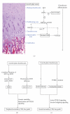
(a) Schematic representations of IHH/PTHLH signaling in the growth plate. The growth plate is composed of chondrocytes at different stages of differentiation, finally leading to longitudinal bone growth. This process is tightly regulated by IHH/PTHLH signaling. IHH, expressed by transition zone chondrocytes, diffuses and binds to Patched (Ptc) in the hypertrophic zone, stimulating PTHLH expression. PTHLH then binds to its receptor in the transition zone and upregulates Bcl-2, which inhibits chondrocyte differentiation and downregulates IHH secretion. EXT gene products play a role in the diffusion of hedgehog proteins and FGF-FGFR interaction. Therefore, defect or absence of EXT genes results in an abnormal IHH diffusion pattern, leading to an osteochondroma. (b) Proposed genetic model for peripheral secondary CHS. Analogous to Knudson's two-hit model, both alleles of an EXT gene are inactivated for osteochondroma formation in both HME and solitary osteochondroma. Genetic instability and reactivation of PTHLH signaling characterizes the malignant transformation of osteochondroma. (c) Proposed genetic model for central secondary CHS. Patients with enchondromatosis infrequently harbor PTHR1 mutation, which disrupts the normal IHH-PTHLH feedback loop, leading to constitutive hedgehog signaling. In most enchondromas, causative genetic or epigenetic changes have not been identified.
Figure 9
Figure 9
Secondary CHS arising in osteochondroma. Note the thickening of cartilage cap (2.5 cm, arrows).
Figure 10
Figure 10
Grade 1 CHS arising in osteochondroma with loss of organized architecture and mild nuclear atypia.
Figure 11
Figure 11
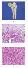
(a) An ill-demarcated lobulating firm mass (8.5 × 4.0 × 4.0 cm) in the proximal femur. Most of the mass which is white to bluish gray is conventional chondrosarcoma. The yellowish gray area in the center (arrows) is dedifferentiated area. (b) The abrupt transition between conventional CHS and dedifferentiated component. (c) The dedifferentiated component consisting of malignant spindle cells without matrix formation (malignant fibrous histiocytoma).
Figure 12
Figure 12
(a) Mesenchymal CHS showing bimorphic pattern, consisting of islands of hyaline cartilage and undifferentiated cells. Note hemangiopericytomatous vascular pattern. (b) The undifferentiated small cells with relatively uniform oval to round nuclei and scanty amount of cytoplasm.
Figure 13
Figure 13
The undifferentiated small cell component in mesenchymal CHS strongly positive for CD99.
Figure 14
Figure 14
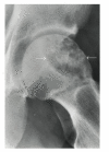
Clear cell CHS located in the epiphysis metaphysis of the proximal femur. The lesion is lytic, slightly expansile, and well delineated from the adjacent normal bone.
Figure 15
Figure 15
Gross finding of clear cell chondrosarcoma in the proximal femur.
Figure 16
Figure 16
(a) Tumor cells with clear cytoplasm between irregular trabeculae of woven bone. (b) Higher magnification of tumor cells with abundant clear or faintly granular cytoplasm and central round nuclei containing occasional prominent nucleoli.

References

    1. Fletcher CDM, Unni KK, Mertens F. World Health Organization Classification of Tumors: Pathology and Genetics of Tumors of Soft Tissue and Bone. Lyon, France: IARC Press; 2002.
    1. Jemal A, Siegel R, Ward E, et al. Cancer statistics, 2006. Ca: A Cancer Journal for Clinicians. 2006;56(2):106–130. - PubMed
    1. Murphey MD, Walker EA, Wilson AJ, Kransdorf MJ, Temple HT, Gannon FH. From the archives of the AFIP: imaging of primary chondrosarcoma: radiologic-pathologic correlation. Radiographics. 2003;23(5):1245–1278. - PubMed
    1. Giuffrida AY, Burgueno JE, Koniaris LG, Gutierrez JC, Duncan R, Scully SP. Chondrosarcoma in the United States (1973 to 2003): an analysis of 2890 cases from the SEER database. Journal of Bone and Joint Surgery. American. 2009;91(5):1063–1072. - PubMed
    1. Murphey MD, Flemming DJ, Boyea SR, Bojescul JA, Sweet DE, Temple HT. Enchondroma versus chondrosarcoma in the appendicular skeleton: differentiating features. Radiographics. 1998;18(5):1213–1237. - PubMed

LinkOut - more resources