. 2011:2011:694136.
doi: 10.1155/2011/694136. Epub 2011 Feb 20.

Osteosarcomagenesis: modeling cancer initiation in the mouse

Affiliations

Osteosarcomagenesis: modeling cancer initiation in the mouse

Kevin B Jones. Sarcoma. 2011.

Abstract

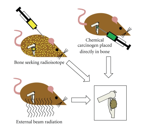
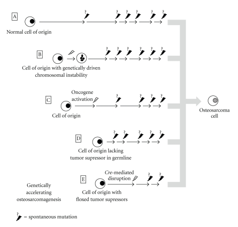
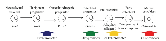
Osteosarcoma remains a deadly malignancy afflicting adolescents and young adults. The lack of a precursor and the panoply of genetic aberrations present in identified osteosarcomas makes study of its initiation difficult. A number of candidate hypotheses have been tested in the mouse, a species with a higher background incidence of osteosarcoma. Chemical carcinogens, external beam radiation, and bone-seeking heavy metal radioisotopes have all proven to be osteosarcomagenic in wild-type mice. A number of oncogenes, introduced via integrating viruses or aberrantly activated from heritable genetic loci, participate in and can individually drive osteosarcomagenesis. Germline and conditional gene ablations in the form of some but not all aneuploidy-inducing genes, conventional tumor suppressors, and factors that function normally in mesenchymal differentiation have also proven osteosarcomagenic, especially in combinations that silence the Rb1 and p53 pathways. This paper reviews the rich history of mouse models of osteosarcomagenesis, what they have taught us about the human disease, and what future mouse experiments yet promise to teach.


Figures

Figure 1
Figure 1
Osteosarcomagenesis can be induced in genetically wild-type mice through a variety of means. Most heavy metal radioisotopes will naturally home to ossifying bone matrix in the metabolically active metaphyses of long bones, emitting their radiation locally after embedding. External beam radiation in varied forms has also proven successfully osteosarcomagenic. Although a few chemical carcinogens administered systemically by oral or intravenous application in mice have proven sufficient, most osteosarcomagenic chemical compounds have been orthotopically implanted in the tibia or femur.
Figure 2
Figure 2
Genetically wild-type cells can naturally accrue sufficient mutations to initiate osteosarcomagenesis in mice as evidenced by the background incidence of osteosarcomas a little under 5 percent in most strains (A). Genetically induced aneuploidy alone, in most of its forms, is inefficient in osteosarcomagenesis (B). Expression of oncogenes as transgenes using native promoters, introduced via insertional viral vectors or unmasked by radiation, can lead to benign and malignant bone neoplasia in mice (C). Inherited germline deletion of tumor suppressors in heterozygosity or homozygosity or generation of mouse chimeras of cells with and without such deletions to avoid serious developmental defects have proven the efficiency of many gene inactivations in osteosarcomagenesis (D). Another means by which severe developmental phenotypes may be eschewed or potential cells of origin tested is by the use of conditional oncogene activation or conditional tumor suppressor ablation, specified either temporally or, by tissue, spatially (E); combinations of tumor suppressor deletions, possible in conditional ablation methods, have been very efficient at driving osteosarcomagenesis.
Figure 3
Figure 3
Osteosarcomagenesis has been tested from a range of cells of origin along the differentiation pathway of osteoblasts. Cre-recombinase expression driven from the Prx1, osterix, and collagen 1a1 promoters has generated osteosarcomas in mice when used for the conditional inactivation of p53 with or without Rb1. The osteocalcin promoter, defining a late stage of osteoblast differentiation usually not expressed in osteosarcoma cells directly, has proven sufficient for osteosarcomagenesis, when used to drive SV40 large and small T antigens.

References

    1. Damron TA, Ward WG, Stewart A. Osteosarcoma, chondrosarcoma, and Ewing’s sarcoma: national cancer data base report. Clinical Orthopaedics and Related Research. 2007;(459):40–47. - PubMed
    1. Unni KK, Dahlin DC. Osteosarcoma: pathology and classification. Seminars in Roentgenology. 1989;24(3):143–152. - PubMed
    1. Marina N, Gebhardt M, Teot L, Gorlick R. Biology and therapeutic advances for pediatric osteosarcoma. Oncologist. 2004;9(4):422–441. - PubMed
    1. Kurt AM, Unni KK, McLeod RA, Pritchard DJ. Low-grade intraosseous osteosarcoma. Cancer. 1990;65(6):1418–1428. - PubMed
    1. Pybus FC, Miller EW. Spontaneous bone tumours of mice. American Journal of Cancer. 1938;33(98):p. 111.