. 2011 Feb 20:2011:767230.
doi: 10.4061/2011/767230.

Mitochondrial fusion/fission, transport and autophagy in Parkinson's disease: when mitochondria get nasty

Affiliations

Mitochondrial fusion/fission, transport and autophagy in Parkinson's disease: when mitochondria get nasty

Daniela M Arduíno et al. Parkinsons Dis. .

Abstract

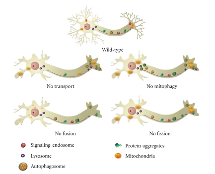
Understanding the molecular basis of Parkinson's disease (PD) has proven to be a major challenge in the field of neurodegenerative diseases. Although several hypotheses have been proposed to explain the molecular mechanisms underlying the pathogenesis of PD, a growing body of evidence has highlighted the role of mitochondrial dysfunction and the disruption of the mechanisms of mitochondrial dynamics in PD and other parkinsonian disorders. In this paper, we comment on the recent advances in how changes in the mitochondrial function and mitochondrial dynamics (fusion/fission, transport, and clearance) contribute to neurodegeneration, specifically focusing on PD. We also evaluate the current controversies in those issues and discuss the role of fusion/fission dynamics in the mitochondrial lifecycle and maintenance. We propose that cellular demise and neurodegeneration in PD are due to the interplay between mitochondrial dysfunction, mitochondrial trafficking disruption, and impaired autophagic clearance.


Figures

Figure 1
Figure 1
Potential susceptibility of neurons to mitochondrial dysfunction and impaired mitochondrial turnover. (Wild type) In healthy neurons, mitochondria, prosurvival signals associated to signaling endosomes, and autophagomes enclosing damaged organelles or protein aggregates are able to travel long distances from the cell periphery to perinuclear region in the cell body, where most lysosomes are concentrated. (No transport) Disruption of microtubule network and subsequent defects on retrograde transport prevent the proper distribution of mitochondria and the efficient transport of autophagy substrates towards lysosomes for degradation, which can lead to defects in energy supply and cargos clearance by autophagy. (No mitophagy) Blockage of autophagic activity seems to be responsible for the accumulation of damaged mitochondria, toxic protein products, aggregates, and leaking autophagic vesicles, all of which have a negative effect on neuronal functioning and survival, precipitating the “dying-back”-type of axonal degeneration. (No fusion) The absence of mitochondrial fusion may result in an accumulation of damaged mitochondria or decreased healthy mitochondria at the nerve terminal. Mitochondria secondarily have defects in motility that prevent proper distribution within the axon and in the periphery. (No fission) In the absence of mitochondrial fission, most of the mitochondrial population is extensively long and interconnected, and a subset shows ultrastructural defects and dysfunction. The large mitochondria clusters within dendrites are not efficiently transported and/or engulfed by autophagosomes towards cell body for lysosomal degradation. (MT: microtubule tracks oriented along the axon with plus (+) ends distal and (−) ends proximal to the cell body).
Figure 2
Figure 2
Rationale for the contribution of mitochondrial dysfunction to synaptic degeneration in sporadic PD. Mitochondrial dysfunction induced by a complex I defect leads to alterations in mitochondria-dependent metabolism (reduced ATP levels and decreased mitochondrial inner membrane potential). This bioenergetic failure seems to potentiate microtubule network breakdown. Subsequently, when the dynamics and functional integrity of microtubules are compromised, changes in anterograde and retrograde flux along the axon can be impaired. Cargos that are actively transported along the axon include mitochondria, autophagosomes, and proteins. Moreover, a decrease in mitochondrial membrane potential deregulates calcium homeostasis, which leads to the overactivation of calpains. Calpains are key regulators of mitochondrial fusion, since they impair Opa1 proper function. Alterations in fusion/fission events promote mitochondrial enlargement, which can impair their removal by mitophagy. Indeed, the accumulation of protein aggregates, autophagosomes, and enlarged deficient mitochondria in presynaptic termini is observed at early stages of PD. Our hypothesis implies that mitochondrial metabolism impairment could be responsible for synaptic degeneration in PD. (*Indicates that mutated or overexpressed α-synuclein could induce mitochondrial dysfunction and that loss-of-function of Parkin or/and PINK1 can deregulate mitochondrial mitophagy.)

Similar articles

Cited by

References

    1. Forno LS. Neuropathology of Parkinson’s disease. Journal of Neuropathology and Experimental Neurology. 1996;55(3):259–272. - PubMed
    1. Banerjee R, Starkov AA, Beal MF, Thomas B. Mitochondrial dysfunction in the limelight of Parkinson’s disease pathogenesis. Biochimica et Biophysica Acta. 2009;1792(7):651–663. - PMC - PubMed
    1. Beal MF. Mitochondria, oxidative damage, and inflammation in Parkinson’s disease. Annals of the New York Academy of Sciences. 2003;991:120–131. - PubMed
    1. Schapira AH. Mitochondria in the aetiology and pathogenesis of Parkinson’s disease. The Lancet Neurology. 2008;7(1):97–109. - PubMed
    1. Cardoso SM, Esteves AR, Arduíno DM, Domingues AF, Oliveira CR. The crucial role of mitochondria in Parkinson’s disease. Recent Research Developments in Neuroscience. 2009;3:43–84.

LinkOut - more resources