

Oligodendrocyte fate after spinal cord injury
- PMID: 21404073
- PMCID: PMC3101831
- DOI: 10.1007/s13311-011-0033-5
Oligodendrocyte fate after spinal cord injury
Abstract
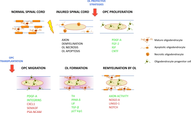
Oligodendrocytes (OLs) are particularly susceptible to the toxicity of the acute lesion environment after spinal cord injury (SCI). They undergo both necrosis and apoptosis acutely, with apoptosis continuing at chronic time points. Loss of OLs causes demyelination and impairs axon function and survival. In parallel, a rapid and protracted OL progenitor cell proliferative response occurs, especially at the lesion borders. Proliferating and migrating OL progenitor cells differentiate into myelinating OLs, which remyelinate demyelinated axons starting at 2 weeks post-injury. The progression of OL lineage cells into mature OLs in the adult after injury recapitulates development to some degree, owing to the plethora of factors within the injury milieu. Although robust, this endogenous oligogenic response is insufficient against OL loss and demyelination. First, in this review we analyze the major spatial-temporal mechanisms of OL loss, replacement, and myelination, with the purpose of highlighting potential areas of intervention after SCI. We then discuss studies on OL protection and replacement. Growth factors have been used both to boost the endogenous progenitor response, and in conjunction with progenitor transplantation to facilitate survival and OL fate. Considerable progress has been made with embryonic stem cell-derived cells and adult neural progenitor cells. For therapies targeting oligogenesis to be successful, endogenous responses and the effects of the acute and chronic lesion environment on OL lineage cells must be understood in detail, and in relation, the optimal therapeutic window for such strategies must also be determined.
Figures
References
-
- Profyris C, Cheema SS, Zang D, Azari MF, Boyle K, Petratos S. Degenerative and regenerative mechanisms governing spinal cord injury. Neurobiol Dis. 2004;15:415–436. - PubMed
-
- Grossman SD, Rosenberg LJ, Wrathall JR. Temporal-spatial pattern of acute neuronal and glial loss after spinal cord contusion. Exp Neurol. 2001;168:273–282. - PubMed
-
- Crowe MJ, Bresnahan JC, Shuman SL, Masters JN, Beattie MS. Apoptosis and delayed degeneration after spinal cord injury in rats and monkeys. Nat Med. 1997;3:73–76. - PubMed
-
- Casha S, Yu WR, Fehlings MG. Oligodendroglial apoptosis occurs along degenerating axons and is associated with FAS and p75 expression following spinal cord injury in the rat. Neuroscience. 2001;103:203–218. - PubMed
Publication types
MeSH terms
LinkOut - more resources
Full Text Sources
Other Literature Sources
Medical
