Review
. 2011 May;20(5):759-72.
doi: 10.1002/pro.613. Epub 2011 Apr 8.

Structural biology of Gram-positive bacterial adhesins

Affiliations
Review

Structural biology of Gram-positive bacterial adhesins

Krishnan Vengadesan et al. Protein Sci. 2011 May.

Abstract

The structural biology of Gram-positive cell surface adhesins is an emerging field of research, whereas Gram-negative pilus assembly and anchoring have been extensively investigated and are well understood. Gram-positive surface proteins known as MSCRAMMs (microbial surface components recognizing adhesive matrix molecules) and individual proteins that assemble into long, hair-like organelles known as pili have similar features at the primary sequence level as well as at the tertiary structural level. Some of these conserved features are essential for their transportation from the cytoplasm and for cell wall anchoring. More importantly, the MSCRAMMs and the individual pilins are assembled with building blocks that are variants of structural modules used for human immunoglobulins. MSCRAMMs target the host's extracellular matrix proteins, such as collagen, fibrinogen, and fibronectin, and they have received considerable attention from structural biologists in the last decade, who have primarily been interested in understanding their interactions with host tissue. The recent focus is on the newly discovered pili of Gram-positive bacteria, and in this review, we highlight the advances in understanding of the individual pilus constituents and their associations and stress the similarities between the individual pilins and surface proteins.


Figures

Figure 1
Figure 1
The domain organization for Gram-positive adhesins. (a) Common structural motifs present in the MSCRAMMs and pilin components. An N-terminal signal sequence (S) followed by a varying number of non-repetitive and repetitive regions suitable for various functions (ligand binding, projecting binding region/stalking, etc.) and C-terminal region for cell wall anchoring that includes a cell wall sorting region (W) containing the LPXTG motif, the membrane-spanning hydrophobic domain (M) and the cytoplasmic positively charged C-terminal tail (C). (b) The organization of three distinct MSCRAMMs. Collagen-binding (CNA/ACE), fibrinogen-binding (ClfA/B, SdrG), and fibronectin-binding (FnbpA/B) MSCRAMMs. In addition to common structural motifs (S, W, M, and C), most adhesins carry ligand-binding domains (N1, N2, N3, …) in the N-terminal half (A-region) and other structural domains that facilitate the extension of A-regions away from the cell surface and unknown functions in the C-terminal half (B-region). Exceptions to this rule are the bifunctional FnbpA/B MSCRAMMs, which display Fn-binding activity at the C-terminal repeats (D1–D4) and fibrinogen-binding activity in the N-terminal half. CNA and ACE are highly homologous; however, the ACE is composed of only two subdomains (N1 and N2) but exhibits equal affinity to the three domain (N1, N2, and N3) CNA. (c) The organization of pilins. In addition to common structural motifs (S, W, M, and C), the pilins carry a varying number of IgG-like domains (often) that makeup pilus shaft and for adhesion. Several major pilins contain conserved pilin motif (WxxxVxVYPK) for covalent polymerization that is catalyzed by an enzyme sortase. The tip pilins contain a vWFA domain, which is flanked by IgG-like domains for adhesion. The base pilin likely contains a lysine in the C-terminal half or in the domain linker for their incorporation into the pilus shaft at the base, and a proline-rich C-terminal tail for cell wall anchoring.
Figure 2
Figure 2
Topology and structure of the IgG constant domain and its variant DEv-IgG, IgG-rev, seen in the MSCRAMMs and pilins. The core β-strands are labeled A–G in a rainbow style color (red to violet). (a) IgG constant domain contains a four-stranded β-sheet I (A, B, E, D) on one side of the barrel and a three-stranded β-sheet II (C, F, G) on the other side. (b) The DEv-IgG fold is similar to IgG-C fold but the additional secondary structural elements (gray) are present between the strands D and E. A possible intra-domain isopeptide bond between A and F strands is shown by a block line. The DEv-IgG fold is first observed in the domains of CNA A-region, hence called as cnaA-type (c) The IgG-rev fold contains a four- (DAGF) and three- (EBC) or three- (DAG) and four- (FEBC) stranded β-sheets similar to IgG-C fold but their arrangements are different. A possible intra-domain isopeptide bond between A and G strands is shown by a block line. The IgG-rev fold is first observed in domains of CNA B repeats, and sometimes described as cnaB-type. [Color figure can be viewed in the online issue, which is available at wileyonlinelibrary.com.]
Figure 3
Figure 3
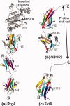
Ribbon representation of minor pilin crystal structures. The IgG-like domains are shown in rainbow style as in Figure 2. The intra-domain isopeptide bonds are shown in sticks. (a) Tip pilin RrgA of S. pneumoniae display two domains of IgG-rev fold (N1 and N4), a DEv-IgG fold domain (N2) and a domain of vWFA fold (N3) with a metal-ion-dependent adhesion site (MIDAS) and two inserted regions. The N2 and N4 domains contain a D- and E-type isopeptide bonds, respectively. (b) Minor pilin GBS52 of S. agalactiae contains two domains (N1 and N2) of IgG-rev fold. The N2 domain has an E-type isopeptide bond and proline-rich C-terminal tail. A lysine (K148) from a pilin-like motif (IYPK) in the domain linker was suggested for its incorporation into the pilus shaft. (c) Minor FctB of S. pyogenes is made of single domain of IgG-rev fold. A lysine (K110) in the Ω-loop was suggested for it incorporation into the pilus shaft. [Color figure can be viewed in the online issue, which is available at wileyonlinelibrary.com.]
Figure 4
Figure 4
Structures of major pilins. The isopeptide bond between the Lys and Asn residues and the conserved proximal Glu residue are shown by sticks. (a) N2 and N3 domains of GBS80 from S. agalactiae. N2 is DEv-IgG fold with D-type isopeptide bond, and two high-affinity metal-binding sites, whereas N3 is an IgG-rev fold with E-type isopeptide bond. (b) Spy0128 from S. pyogenes is made of two IgG-rev fold domains, each with an E-type isopeptide bond. A lysine (K161) in the Ω-loop was suggested for pilus polymerization. (c) SpaA from C. diphtheriae shows three IgG-like domains (N1, N2, and N3). N1 and N3 exhibits IgG-rev folds, whereas N2 exhibits DEv-IgG fold, and hosts a metal-binding site and D-type isopeptide bond. The N3 contains an E-type isopeptide bond while N1 does not. The pilin motif lysine (K190) is seen at N1-N2 domain linker. (d) N2, N3, and N4 domains of BcpA from B. cereus. The N2 and N4 exhibit IgG-rev fold while N3 is of DEv-IgG fold. (e) N2, N3, and N4 domains of RrgB from S. pneumonia. The N2 domain display a DEv-IgG fold, whereas N3 and N4 domains show IgG-rev fold with respective isopeptide bonds. The N2 and N3 domains are packed parallel, and N3 domain is inserted into N2 domain. [Color figure can be viewed in the online issue, which is available at wileyonlinelibrary.com.]
Figure 5
Figure 5
Crystal structures of MSCRAMMs as apo- and in complex with ligands. (a) Ribbon and surface representation collagen-binding domains (N1 and N2) of CNA. In the ribbon diagram, the N1 and N2 domains are shown in rainbow colors, whereas in the surface diagram they are shown in light green and cyan, respectively. A long flexible linker (in pink) connects the N1 and N2 domains. The C-terminus of the N2 domain extends toward and into the N1 domain and forms a β-strand (in magenta) that complements one of the β-sheets of the N1 domain. The linker is partially disordered in the apo structure and it is modeled to show the hole between the N1 and N2 domains. (b) Stereo representation of CNAN1N2 in complex with collagen-like triple helical peptide. The three chains of the collagen peptide (in the surface representation) are leading (L) (coral), middle (M) (light yellow), and trailing (T) (light blue), as viewed from their N-termini. Two CNA molecules [one in the ribbon and another in surface representation and colored as in Fig. 5(a)] interact with the collagen peptide in an identical manner. The leading and trailing chains of the peptide interact with the N2 domain and the middle chain with N1. The N1-N2 linker covers the leading and trailing chains and holds the rope-like ligand in place. (c) Ribbon representation of N2 and N3 domains of SdrG. N2 and N3 domains are shown in rainbow colors. The N2-N3 linker is represented in pink and the C-terminal is disordered in the apo structure. (d) Surface representation of SdrGN2N3 in complex with the fibrinogen peptide. The N2 domain is in light green, N3 is in cyan and the inter-domain linker is in pink, whereas the Fg peptide is shown as a ribbon in red. The C-terminal latch region is shown in magenta. [Color figure can be viewed in the online issue, which is available at wileyonlinelibrary.com.]

Similar articles

Cited by

References

    1. Kline KA, Dodson KW, Caparon MG, Hultgren SJ. A tale of two pili: assembly and function of pili in bacteria. Trends Microbiol. 2010;18:224–232. - PMC - PubMed
    1. Sauer FG, Mulvey MA, Schilling JD, Martinez JJ, Hultgren SJ. Bacterial pili: molecular mechanisms of pathogenesis. Curr Opin Microbiol. 2000;3:65–72. - PubMed
    1. Waksman G, Hultgren SJ. Structural biology of the chaperone-usher pathway of pilus biogenesis. Nat Rev Microbiol. 2009;7:765–774. - PMC - PubMed
    1. Choudhury D, Thompson A, Stojanoff V, Langermann S, Pinkner J, Hultgren SJ, Knight SD. X-ray structure of the FimC-FimH chaperone-adhesin complex from uropathogenic Escherichia coli. Science. 1999;285:1061–1066. - PubMed
    1. Scott JR, Zahner D. Pili with strong attachments: Gram-positive bacteria do it differently. Mol Microbiol. 2006;62:320–330. - PubMed

MeSH terms

Substances

LinkOut - more resources