

doi: 10.1103/PhysRevLett.106.034301.
Epub 2011 Jan 18.
Blood vessel deformations on microsecond time scales by ultrasonic cavitation
Affiliations
- PMID: 21405276
- PMCID: PMC3087441
- DOI: 10.1103/PhysRevLett.106.034301
Item in Clipboard
Blood vessel deformations on microsecond time scales by ultrasonic cavitation
Phys Rev Lett.
.
Abstract
Transient interactions among ultrasound, microbubbles, and microvessels were studied using high-speed photomicrography. We observed liquid jets, vessel distention (motion outward against the surrounding tissue), and vessel invagination (motion inward toward the lumen). Contrary to current paradigms, liquid jets were directed away from the nearest vessel wall and invagination exceeded distention. These observations provide insight into the mechanics of bubble-vessel interactions, which appear to depend qualitatively upon the mechanical properties of biological tissues.
Figures
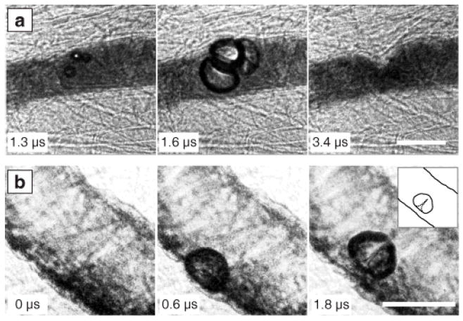
Characteristics of observed bubble-vessel interactions. (a) A group of bubbles distends the vessel wall (middle); subsequent invagination (right) appears localized and markedly larger than the distention. (b) A bubble distends the vessel wall (middle) and then forms a liquid jet directed away from this wall (right, with inset showing a sketch for clarity). Complete image sequences are shown in Movies S1 and S2 of the supplementary material [20]. In all the figures, time stamps indicate the time after arrival of the start of the ultrasound pulse, and scale bars represent 50 μm.
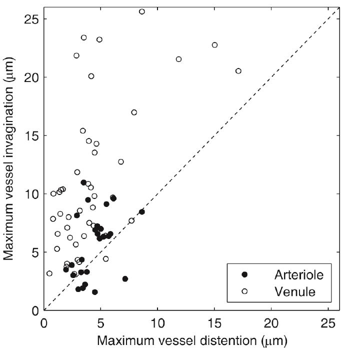
Comparison of maximum vessel invagination and distention. Each data point represents observed changes in vessel diameter for an interaction between an isolated or dominant target bubble and a vessel. Observations include microvessels with diameters between 10 and 100 μm and ultrasound pulses with peak negative pressures (PNP) between 0.8 and 7.2 MPa. In 60 out of 70 cases, the data fall above the dashed line, demonstrating that invagination typically exceeded distention.
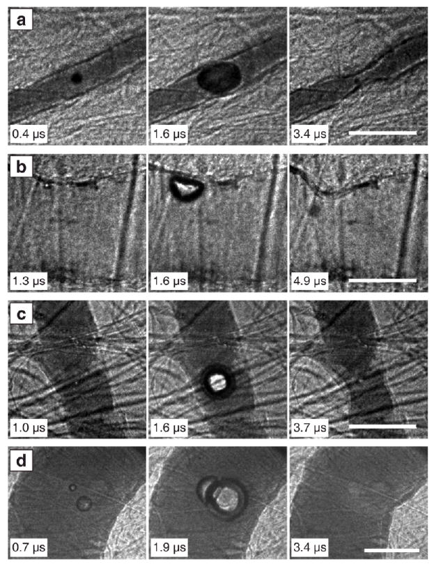
Image sequences to illustrate types of vessel invagination. In a and b, localized vessel invagination was observed when the bubble contacted the vessel wall. In c and d, the bubble did not contact the vessel wall, but still induced local vessel invagination. (a) PNP = 1.5 MPa, vessel diameter = 22 μm. (b) PNP = 4.0 MPa, vessel diameter = 71 μm. (c) PNP = 0.9 MPa, vessel diameter = 42 μm. (d) PNP = 7.2 MPa, vessel diameter = 100 μm. Complete image sequences corresponding to a–d are shown in Movies S3–S6 of the supplementary material [20].
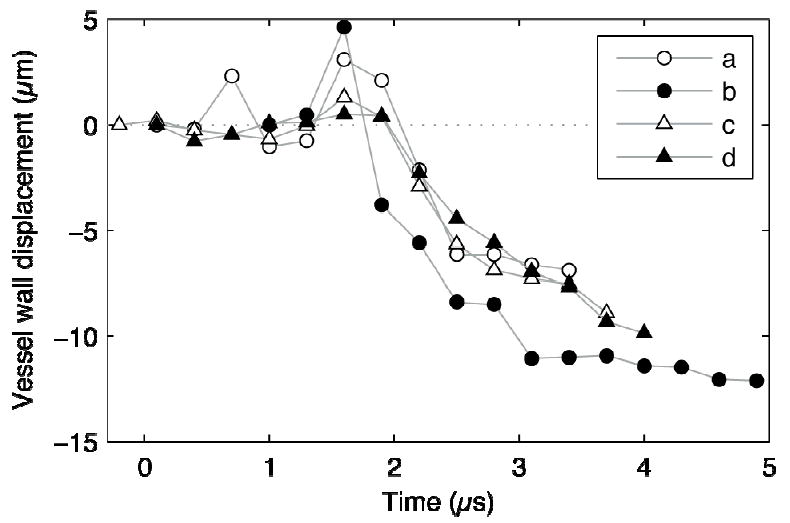
Measurements of radial displacements of the vessel wall at the point closest to the bubble for Figs. 2a–d. Each marker denotes a measurement from a single image frame. Deflections toward the lumen were defined to be negative. For each of these sequences, vessel invagination exceeded distention by a significant margin. The observed invaginations occurred after bubbles collapsed (at about 2 μs in the plot) and persisted even after bubbles rebounded.
References
Publication types
MeSH terms
Grants and funding
LinkOut - more resources
Full Text Sources