Review
. 2011 Apr 1;435(1):1-16.
doi: 10.1042/BJ20100965.

Proteases as therapeutics

Affiliations
Review

Proteases as therapeutics

Charles S Craik et al. Biochem J. .

Abstract

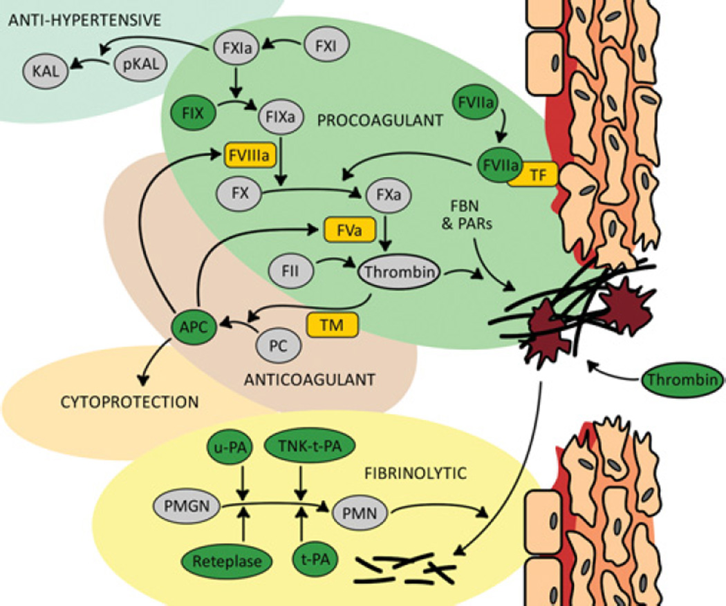
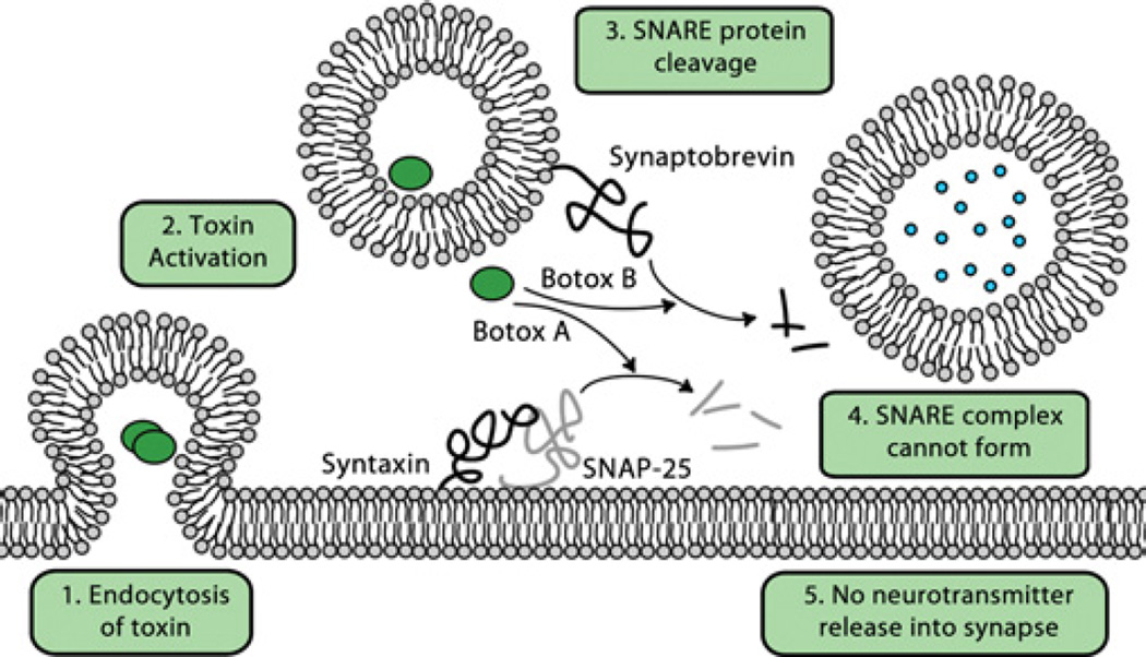
Proteases are an expanding class of drugs that hold great promise. The U.S. FDA (Food and Drug Administration) has approved 12 protease therapies, and a number of next generation or completely new proteases are in clinical development. Although they are a well-recognized class of targets for inhibitors, proteases themselves have not typically been considered as a drug class despite their application in the clinic over the last several decades; initially as plasma fractions and later as purified products. Although the predominant use of proteases has been in treating cardiovascular disease, they are also emerging as useful agents in the treatment of sepsis, digestive disorders, inflammation, cystic fibrosis, retinal disorders, psoriasis and other diseases. In the present review, we outline the history of proteases as therapeutics, provide an overview of their current clinical application, and describe several approaches to improve and expand their clinical application. Undoubtedly, our ability to harness proteolysis for disease treatment will increase with our understanding of protease biology and the molecular mechanisms responsible. New technologies for rationally engineering proteases, as well as improved delivery options, will expand greatly the potential applications of these enzymes. The recognition that proteases are, in fact, an established class of safe and efficacious drugs will stimulate investigation of additional therapeutic applications for these enzymes. Proteases therefore have a bright future as a distinct therapeutic class with diverse clinical applications.


Figures

Figure 1
Figure 1. Protease therapeutics applied successfully for procoagulant and fibrinolytic indications
Proteolytic cascades are responsible for the formation and dissolution of blood clots and therefore they can be used individually for therapeutic benefit. Eight proteases (shown in dark green circles) have been approved for clinical use. The use of proteases for benefits outside of clotting has begun to emerge from a deeper understanding of protease biology and better delivery technologies. For example, the anticoagulant APC may be modified for its anti-inflammatory effect whereas tissue prekallikrein (pKAL) is a target for gene therapy owing to its anti-hypertensive potential. Protein cofactors are represented by rectangles with rounded edges. FBN, fibronectin; FII, prothrombin; KAL, kallikrein; PC, protein C; PMGN, plasminogen; PMN, plasmin; TNKase, tenecteplase.
Figure 2
Figure 2. Mechanism of botulinum toxin action
Endocytosis leads to activation of the toxin and separation into its heavy and light chains. The light-chain protease transports to the cytoplasm where it degrades one or more proteins involved in SNARE-mediated vesicle transport. The downstream effect of these proteolytic events is a diminished release of acetylcholine and neurotransmission. Botulinum toxin type A (Botox A) and botulinum toxin type B (Botox B) have different substrates in the synaptic fusion complex. Botox A cleaves synaptosomal-associated protein of 25 kDa (SNAP-25), whereas Botox B inactivates synaptobrevin, and both are key members of the SNARE complex. An animated version of this Figure is available at http://www.BiochemJ.org/bj/435/bj4350001add.htm
Figure 3
Figure 3. The trypsin fold is the most common protease fold found in the genomes of higher organisms [186] and is the most commonly scaffold of existing therapeutic proteases
Shown is the structure of the protease domain t-PA, which like other members of the family, contains two six-stranded Greek key β-barrels lying on top of and perpendicular to one another with the active-site cleft between them [187]. Eight loops surround the catalytic triad (green) and the primary specificity pocket (red) of which five have been particularly useful targets for protein engineering. The loops above the active-site cleft near the catalytic triad (blue) make direct contacts with the substrate near the scissile bond and have been manipulated widely [188]. For reference a tripeptide inhibitor is shown in orange. The positions outside of the active-site cleft (yellow) have been engineered to restrict the interaction with macromolecular substrates and inhibitors [189]. For example, four successive alanine residue substitutions in Loop A were used to restrict the interaction of t-PA with PAI-1 [190]. Alterations within the active-site cleft have also been made to modify the properties of the protease. In thrombin such changes have been used to shift the protein to an anticoagulant with limited procoagulant functionality [64,191]. The presence or absence of protein domains attached to the protease domain can be engineered for improved therapeutic efficacy. The loops are labelled and numbered according to [192]: Loop A, residues 34–41; Loop B, residues 56–64; Loop C, residues 94–103; Loop D, residues 143–151; and Loop E, residues 74–80 in the chymotrypsin numbering system.

References

    1. Rodriguez D, Morrison CJ, Overall CM. Matrix metalloproteinases: what do they not do? New substrates and biological roles identified by murine models and proteomics. Biochim. Biophys. Acta. 2010;1803:39–54. - PubMed
    1. Duffy MJ, McGowan PM, Gallagher WM. Cancer invasion and metastasis: changing views. J. Pathol. 2008;214:283–293. - PubMed
    1. Overall CM, Blobel CP. In search of partners: linking extracellular proteases to substrates. Nat. Rev. Mol. Cell Biol. 2007;8:245–257. - PubMed
    1. Puente XS, Sánchez LM, Gutiérrez-Fernández A, Velasco G, López-Otín C. A genomic view of the complexity of mammalian proteolytic systems. Biochem. Soc. Trans. 2005;33:331–334. - PubMed
    1. Puente XS, Lopez-Otin C. A genomic analysis of rat proteases and protease inhibitors. Genome Res. 2004;14:609–622. - PMC - PubMed

MeSH terms