. 2011 Mar 15;71(6):2360-70.
doi: 10.1158/0008-5472.CAN-10-1933.

Histone methyltransferase EZH2 induces Akt-dependent genomic instability and BRCA1 inhibition in breast cancer

Affiliations

Histone methyltransferase EZH2 induces Akt-dependent genomic instability and BRCA1 inhibition in breast cancer

Maria E Gonzalez et al. Cancer Res. .

Abstract

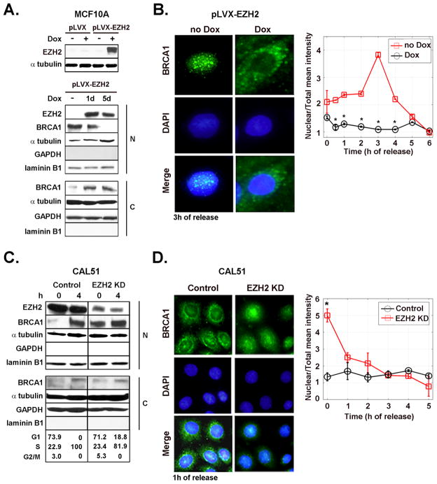
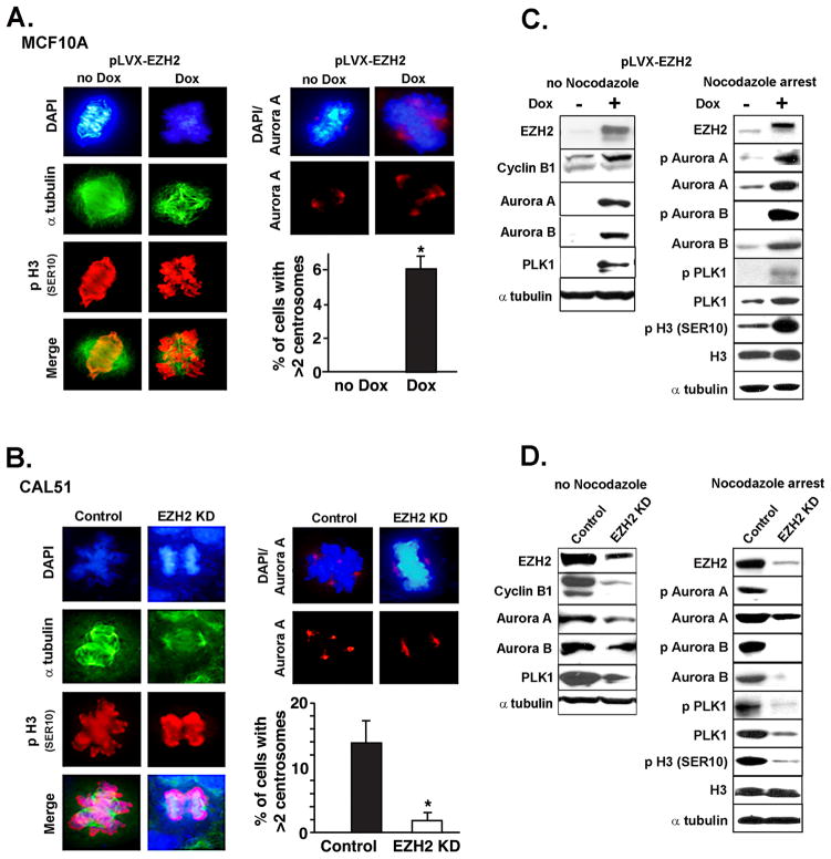
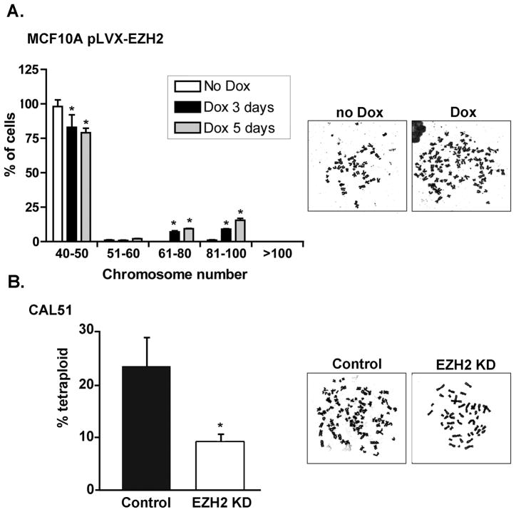
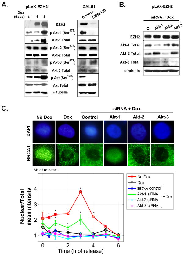
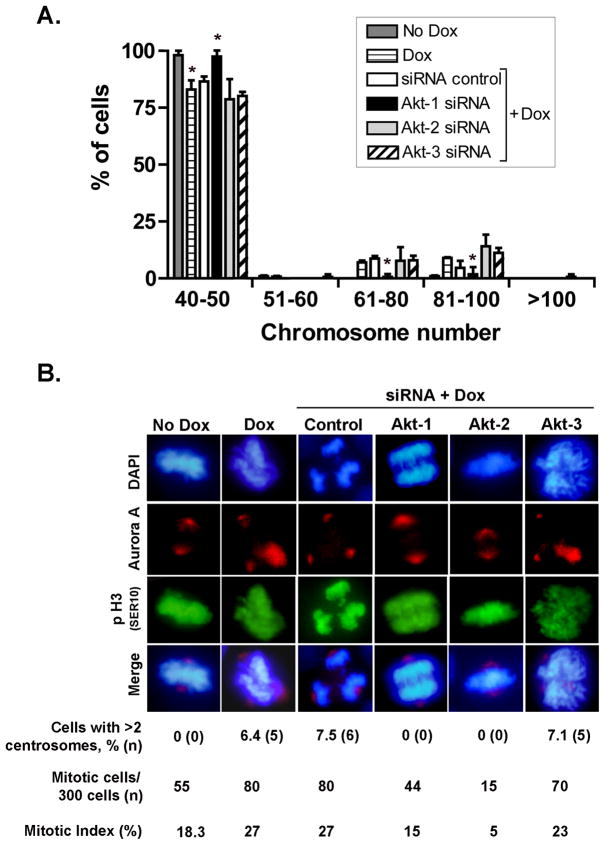
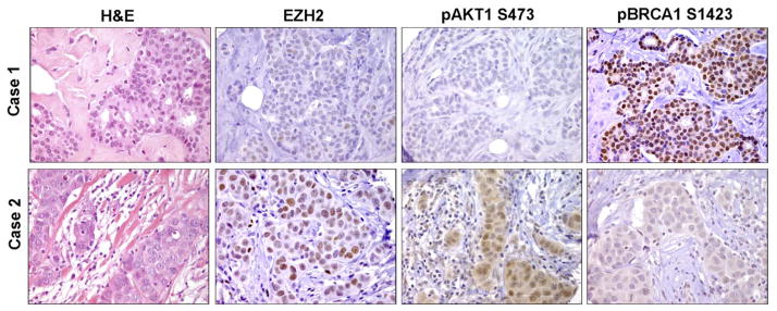
Increased levels of EZH2, a critical regulator of cellular memory, signal the presence of metastasis and poor outcome in breast cancer patients. High levels of EZH2 are associated with nuclear pleomorphism, lack of estrogen receptor expression, and decreased nuclear levels of BRCA1 tumor suppressor protein in invasive breast carcinomas. The mechanism by which EZH2 overexpression promotes the growth of poorly differentiated invasive carcinomas remains to be defined. Here, we show that EZH2 controls the intracellular localization of BRCA1 protein. Conditional doxycycline-induced upregulation of EZH2 in benign mammary epithelial cells results in nuclear export of BRCA1 protein, aberrant mitoses with extra centrosomes, and genomic instability. EZH2 inhibition in CAL51 breast cancer cells induces BRCA1 nuclear localization and rescues defects in ploidy and mitosis. Mechanistically, EZH2 overexpression is sufficient for activation of the phosphoinositide 3-kinase/Akt (PI3K/Akt) pathway specifically through activation of Akt isoform 1. EZH2-induced BRCA1 nuclear export, aneuploidy, and mitotic defects were prevented by treatment with the PI3K inhibitors LY294002 or wortmannin. Targeted inhibition of Akt-1, Akt-2, and Akt-3 isoforms revealed that the EZH2-induced phenotype requires specific activation of Akt-1. The relevance of our studies to human breast cancer is highlighted by the finding that high EZH2 protein levels are associated with upregulated expression of phospho-Akt-1 (Ser473) and decreased nuclear expression of phospho-BRCA1 (Ser1423) in 39% of invasive breast carcinomas. These results enable us to pinpoint one mechanism by which EZH2 regulates BRCA1 expression and genomic stability mediated by the PI3K/Akt-1 pathway.


Figures

Figure 1
Figure 1. EZH2-dependent regulation of BRCA1 intracellular localization
A. Inducible synthesis of EZH2 in MCF10A cells and its effect on BRCA1 protein. Western blot analysis of MCF10A cells transduced with the empty vector (pLVX) and with EZH2-containing vector (pLVX-EZH2). Cells were untreated or treated with Dox (500 ng/ml) to induce EZH2 expression. Underneath is a Western blot of EZH2 and BRCA1 proteins in nuclear and cytoplasmic enriched fractions of pLVX-EZH2 cells untreated or treated with Dox. Laminin B1 and GAPDH confirm nuclear and cytoplasmic enrichment of the fractionated samples, respectively. B. Effect of EZH2 overexpression on the intracellular localization of BRCA1 protein. Representative immunofluorescence images of BRCA1 protein at 3 h after release of G1/S cell cycle block (double thymidine block). The graph shows the mean intensity of BRCA1 protein expression in the nucleus normalized to the total intensity at different times after release from cell cycle block. The percentage of cells in each cell cycle phase after release of cell cycle block for untreated and Dox treated cells was at 0 h: G1 94.84%, S 5.15%, G2/M 0%, and G1 94.09%; S 4.97%, G2/M 0.94%, respectively. At 4 h: G1 78.52%, S 21.02%, G2/M 0.46%, and G1 74.27%, S 25.7%, G2/M 0.03%, respectively. C. Western blot for EZH2 and BRCA1 proteins in CAL51/EZH2 KD and controls at 0 and 4 h after release of cell cycle block. The number of cells in G1, S and M cell cycle phases was obtained by flow cytometry. D. Representative immunofluorescence images of BRCA1 protein at 1 h after release of cell cycle block and quantification of results, as described above. The experiment was repeated for three independent times. Error bars are the standard deviation and * represents p<0.05.
Figure 2
Figure 2. EZH2-mediated induction of abnormal mitosis and Aurora kinase activity
A and B. Immunofluorescence analysis of mitotic MCF10A- pLVX-EZH2 cells untreated or treated with Dox for 3 days (A), and in CAL51/KD and controls (B). p-H3 marks mitotic DNA; Aurora A marks the centrosomes. Bar graphs show the percentage of cells with > 2 centrosomes ± SD. The number of mitotic cells per 300 cells was 59 (19%) and 78 (26%) for untreated and Dox treated cells, respectively. The number of mitotic cells per 300 cells was 198 (66%) and 78 (26%) for control and EZH2 KD cells, respectively. C. Western blot analysis of MCF10A-pLVX-EZH2 cells untreated or treated with Nocodazole (50 ng/ml for 20 h at 80% confluence) to induce mitotic arrest. EZH2 overexpression induces Aurora A and B levels in asynchronized cultures, and upregulates p Aurora A and B, p Plk1, and the Aurora kinase substrate p H3 Ser10 in Nocodazole treated cells. D. Western blot of CAL51 cells untreated or treated with Nocodazole. EZH2 KD in CAL51 cells show the opposite effects than EZH2 overexpression.
Figure 3
Figure 3. EZH2 regulates genomic stability in benign and breast cancer cells
A. MCF10A-pLVX-EZH2 cells untreated or treated with Dox for 3 and 5 days were subjected to Giemsa-stained metaphase spread analysis. While untreated cells are diploid, EZH2 overexpression induced polyploidy (≥51 chromosomes) and increased near tetraploid cells (81–100 chromosomes). Bar graph shows the mean percentage of cells ± SD. B. Metaphase counts in CAL51 control and EZH2 KD cells. For both experiments, 100 metaphases per sample were counted and the assay was performed in triplicate. * p<0.05.
Figure 4
Figure 4. EZH2 requires Akt-1 to regulate the intracellular localization of BRCA1
A. Immunoblots of MCF10A and CAL51 cells show that EZH2 expression regulates Akt-1 protein, while it has no effect on Akt-2 or Akt-3 protein levels. B. Western blot of MCF10A-pLVX-EZH2 cells treated with siRNA control or siRNA-Akt-1, siRNA-Akt-2, or siRNA-Akt-3 and then subjected to Dox for 3 days to induce EZH2 overexpression. Immunoblot shows specific inhibition of Akt isoforms. C. Cells described in B as well as pLVX-EZH2 untreated or treated with Dox were subjected to immunofluorescence analyses of BRCA1 protein at the indicated time points after release from cell cycle block at G1/S. Representative confocal images at 3 h after release from cell cycle block.Greater than 500 cells were counted per slide, in triplicate. Mean intensity of BRCA1 protein in the nucleus was normalized to the total intensity utilizing the ImageJ NIH program. Error bars are the standard deviation. * p<0.05.
Figure 5
Figure 5. Akt-1 is required for EZH2-dependent regulation of mitosis and genomic stability
A. MCF10A-pLVX-EZH2 cells treated with siRNA control or siRNA-Akt-1, siRNA-Akt-2, or siRNA-Akt-3 were subjected to Dox for 3 days to induce EZH2 overexpression. Giemsa-stained metaphase spread analyses show that siRNA-Akt-1 is sufficient to prevent EZH2-induced polyploidy (≥51 chromosomes). In contrast, EZH2 overexpression increased numerical chromosomal alterations in cells treated with siRNA control, siRNA Akt-2, or siRNA Akt-3. Bar graph shows the mean percentage of cells ± SD. B. Immunofluorescence analysis of mitotic MCF10A- pLVX-EZH2 cells treated with siRNA control or siRNA-Akt-1, siRNA-Akt-2, or siRNA-Akt-3 and were then subjected to Dox for 3 days. p-H3 (Ser10) marks mitotic DNA; Aurora A marks the centrosomes, DAPI marks the nuclei. Note that Akt-1 siRNA prevented EZH2-induced mitotic aberrations and the number of cells with extra centrosomes. Akt-2 siRNA caused a 3-fold decrease in the number of mitotic cells independent of EZH2 overexpression (mitotic index for siRNA control/no Dox = 18.3% and for siRNA control/Dox = 27%; compared to mitotic index for siRNA Akt-2/no Dox = 5.6% and for siRNA Akt-2/Dox = 5%). Akt-3 KD was not able to prevent EZH2-induced chromosomal alterations.
Figure 6
Figure 6. EZH2 overexpression is associated with upregulation of pAkt1 and decreased nuclear BRCA1 expression
Human breast cancer tissue samples (n=138) were immunostained for EZH2, pAkt1, and pBRCA1. Representative images of invasive ductal carcinomas.

References

    1. Laible G, Wolf A, Dorn R, et al. Mammalian homologues of the Polycomb-group gene Enhancer of zeste mediate gene silencing in Drosophila heterochromatin and at S. cerevisiae telomeres. Embo J. 1997;16:3219–32. - PMC - PubMed
    1. Satijn DP, Otte AP. Polycomb group protein complexes: do different complexes regulate distinct target genes? Biochim Biophys Acta. 1999;1447:1–16. - PubMed
    1. Ringrose L, Paro R. Epigenetic regulation of cellular memory by the Polycomb and Trithorax group proteins. Annu Rev Genet. 2004;38:413–43. - PubMed
    1. Haupt Y, Alexander WS, Barri G, Klinken SP, Adams JM. Novel zinc finger gene implicated as myc collaborator by retrovirally accelerated lymphomagenesis in E mu-myc transgenic mice. Cell. 1991;65:753–63. - PubMed
    1. Hess JL. MLL: a histone methyltransferase disrupted in leukemia. Trends Mol Med. 2004;10:500–7. - PubMed

Publication types

MeSH terms