. 2011 Mar 15;25(6):646-59.
doi: 10.1101/gad.2004211.

Activation of miR-31 function in already-established metastases elicits metastatic regression

Affiliations

Activation of miR-31 function in already-established metastases elicits metastatic regression

Scott Valastyan et al. Genes Dev. .

Retraction in

Abstract

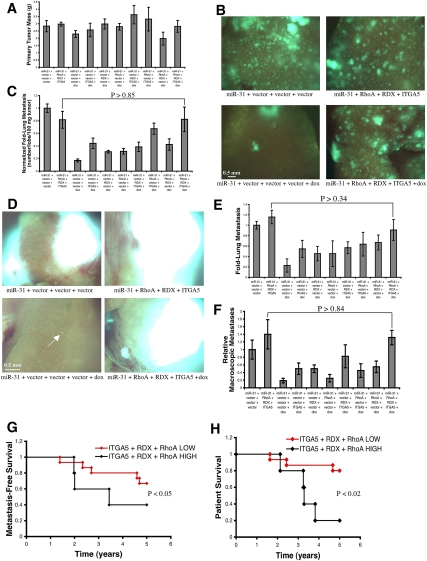
Distant metastases, rather than the primary tumors from which these lesions arise, are responsible for >90% of carcinoma-associated mortality. Many patients already harbor disseminated tumor cells in their bloodstream, bone marrow, and distant organs when they initially present with cancer. Hence, truly effective anti-metastatic therapeutics must impair the proliferation and survival of already-established metastases. Here, we assess the therapeutic potential of acutely expressing the microRNA miR-31 in already-formed breast cancer metastases. Activation of miR-31 in established metastases elicits metastatic regression and prolongs survival. Remarkably, even brief induction of miR-31 in macroscopic pulmonary metastases diminishes metastatic burden. In contrast, acute miR-31 expression fails to affect primary mammary tumor growth. miR-31 triggers metastatic regression in the lungs by eliciting cell cycle arrest and apoptosis; these responses occur specifically in metastases and can be explained by miR-31-mediated suppression of integrin-α5, radixin, and RhoA. Indeed, concomitant re-expression of these three proteins renders already-seeded pulmonary metastases refractory to miR-31-conferred regression. Upon miR-31 activation, Akt-dependent signaling is attenuated and the proapoptotic molecule Bim is induced; these effects occur in a metastasis-specific manner in pulmonary lesions and are abrogated by concurrent re-expression of integrin-α5, radixin, and RhoA. Collectively, these findings raise the possibility that intervention strategies centered on restoring miR-31 function may prove clinically useful for combating metastatic disease.


Figures

Figure 1.
Figure 1.
Acute miR-31 expression drives regression of established spontaneous lung metastases. (A) Overview of the dox-controlled intervention strategy for acute miR-31 expression upon orthotopic implantation of the indicated GFP-labeled 231 cells. (B) Masses of 231 cell primary mammary tumors 56 d after orthotopic injection. n = 5. (C) Hematoxylin and eosin (H&E) staining of primary mammary tumors 56 d subsequent to orthotopic implantation. (D) Quantification of primary mammary tumor local invasion 56 d following orthotopic injection. n = 5. (Asterisks) P < 0.04 relative to reverse tetracycline-controlled transactivator (rtTA)-miR-31 cells (no dox treatment). (E) Fluorescent images of murine lungs to visualize disseminated 231 cells 56 d after orthotopic implantation. (F) Quantification of lung metastatic burden 56 d after orthotopic injection. n = 5. (Asterisks) P < 0.04 relative to rtTA-miR-31 cells (no dox treatment). All error bars represent mean ± SEM.
Figure 2.
Figure 2.
Acute miR-31 expression after primary mammary tumor resection triggers regression of established spontaneous pulmonary metastases. (A) Schematic depicting the dox-controlled intervention strategy for acute miR-31 expression upon orthotopic implantation of the indicated GFP-labeled 231 cells. Primary mammary tumors were surgically resected 26 d after injection. (B) Masses of resected 231 cell primary mammary tumors 26 d subsequent to orthotopic implantation. n = 4. (C) Fluorescent images of murine lungs to visualize disseminated 231 cells 66 d after orthotopic injection. (D) Quantification of metastatic burden in the lungs 66 d after orthotopic implantation. n = 4. (Asterisks) P < 0.004 relative to rtTA-miR-31 cells (no dox treatment). (E) Kaplan-Meier curves depicting survival in this assay. n = 5. (Asterisks) P < 0.05 relative to rtTA-miR-31 cells (no dox treatment); P-value is based on a log-rank test. All error bars represent mean ± SEM.
Figure 3.
Figure 3.
miR-31 activation drives regression of established experimental lung metastases. (A) Overview of the dox-mediated intervention strategy for acute miR-31 expression upon intravenous injection of the indicated GFP-labeled 231 cells via the tail vein. Images document the normal progression of control 231 cells lacking miR-31 in the lungs in this assay. (Arrows) Micrometastases. (B) Fluorescent images of murine lungs to visualize 231 cells 88 d after intravenous implantation. (Arrows) Micrometastases. (C) Quantification of metastatic burden in the lungs 88 d following intravenous implantation. n = 5. (Asterisks) P < 0.05 relative to rtTA-miR-31 cells (no dox treatment). (D) Quantification of the relative prevalence of macroscopic metastases in the lungs 88 d subsequent to intravenous injection. n = 5. (Asterisks) P < 0.05 relative to rtTA-miR-31 cells (no dox treatment). (E) Kaplan-Meier curves depicting survival in this assay. n = 15. (Asterisks) P < 0.05 relative to rtTA-miR-31 cells (no dox treatment); P-value is based on a log-rank test. All error bars represent mean ± SEM.
Figure 4.
Figure 4.
Activation of miR-31 in established pulmonary metastases triggers metastasis-specific cell cycle arrest and apoptosis. (A) 231 cell lung metastases 88 d after intravenous injection via the tail vein, immunohistochemically stained for phosphorylated histone H3 (phospho-H3). (B) Quantification of phospho-H3 staining in pulmonary metastases 88 d post-intravenous implantation. n = 5. (Asterisks) P < 0.03 relative to rtTA-miR-31 cells (no dox treatment). (C) 231 cell lung metastases 88 d after intravenous introduction, immunohistochemically stained for cleaved caspase3. (D) Quantification of cleaved caspase3 staining in pulmonary metastases 88 d following intravenous injection. n = 5. (Asterisks) P < 0.03 relative to rtTA-miR-31 cells (no dox treatment). (E) Quantification of phospho-H3 staining in 231 cell primary mammary tumors 56 d post-orthotopic injection. n = 5. (Asterisks) P < 0.03 relative to rtTA-miR-31 cells (no dox treatment). (F) Quantification of cleaved caspase3 staining in 231 cell primary mammary tumors 56 d subsequent to orthotopic implantation. n = 5. (G) In situ hybridizations for miR-31 (green) in animal-matched 231 cell primary mammary tumors and lung metastases 56 d following orthotopic implantation. (Blue) DAPI counterstain. (H) Quantification of miR-31 staining in animal-matched primary mammary tumors and lung metastases 56 d post-orthotopic injection. n = 5. (Asterisks) P < 0.03 relative to rtTA-miR-31 cells (no dox treatment). All error bars represent mean ± SEM.
Figure 5.
Figure 5.
Suppression of ITGA5, RDX, and RhoA can mediate miR-31-evoked regression of established pulmonary metastases. (A) Masses of 231 cell primary mammary tumors 54 d subsequent to orthotopic injection of the indicated GFP-labeled 231 cells. n = 4. (miR-31) rtTA-miR-31. (B) Fluorescent images of murine lungs to visualize disseminated 231 cells 54 d after orthotopic implantation. (C) Quantification of metastatic burden in the lungs 54 d after orthotopic injection. n = 4. (D) Fluorescent images of murine lungs to visualize 231 cells 92 d after intravenous implantation via the tail vein. (Arrows) Micrometastases. (E) Quantification of metastatic burden in the lungs 92 d following intravenous implantation. n = 5. (F) Quantification of the relative prevalence of macroscopic metastases in the lungs 92 d subsequent to intravenous injection. n = 5. (G) Kaplan-Meier curves for 295 human primary breast tumors depicting 5-yr metastasis-free survival, stratified based on coordinate differential expression of ITGA5, RDX, and RhoA; P-value based on a log-rank test. (H) Kaplan-Meier 5-yr survival curves for 295 breast cancer patients, stratified based on coordinate differential expression of ITGA5, RDX, and RhoA; P-value is based on a log-rank test. All error bars represent mean ± SEM.
Figure 6.
Figure 6.
ITGA5, RDX, and RhoA can control metastasis-specific cell cycle arrest and apoptosis evoked by miR-31 activation in established lung metastases. (A) 231 cell lung metastases 54 d after orthotopic injection, immunohistochemically stained for phospho-H3. (miR-31) rtTA-miR-31. (B) Quantification of phospho-H3 staining in pulmonary metastases 54 d subsequent to orthotopic implantation. n = 5. (C) 231 cell lung metastases 54 d post-orthotopic introduction, immunohistochemically stained for cleaved caspase3. (D) Quantification of cleaved caspase3 staining in pulmonary metastases 54 d following orthotopic injection. n = 5. (E) Quantification of miR-31 staining, as achieved by in situ hybridizations, in animal-matched primary mammary tumors and lung metastases 54 d after orthotopic injection. n = 5. All error bars represent mean ± SEM.
Figure 7.
Figure 7.
The Akt and Bim pathways are altered by miR-31, ITGA5, RDX, and RhoA in a metastasis-specific manner in established pulmonary metastases. (A) Quantification of phosphorylated Akt (phospho-Akt) staining in 231 cell pulmonary metastases 56 d following orthotopic implantation. n = 5. (Asterisks) P < 0.02 relative to rtTA-miR-31 cells (no dox treatment). (B) Quantification of phospho-Akt staining in 231 cell primary mammary tumors 56 d post-orthotopic injection. n = 5. (C) Quantification of Bim immunohistochemical staining in 231 cell pulmonary metastases 56 d following orthotopic implantation. n = 5. (Asterisks) P < 0.02 relative to rtTA-miR-31 cells (no dox treatment). (D) Quantification of Bim immunohistochemical staining in 231 cell primary mammary tumors 56 d post-orthotopic injection. n = 5. (E) 231 cell lung metastases 54 d subsequent to orthotopic introduction, immunohistochemically stained for phospho-Akt. (F) Quantification of phospho-Akt staining in 231 cell pulmonary metastases 54 d following orthotopic implantation. n = 5. (G) 231 cell lung metastases 54 d after orthotopic introduction, immunohistochemically stained for Bim. (H) Quantification of Bim immunohistochemical staining in 231 cell pulmonary metastases 54 d post-orthotopic injection. n = 5. All error bars represent mean ± SEM.

References

    1. Chen L, Yang S, Jakoncic J, Zhang JJ, Huang X 2010. Migrastatin analogues target fascin to block tumour metastasis. Nature 464: 1062–1066 - PMC - PubMed
    1. Coussens LM, Fingleton B, Matrisian LM 2002. Matrix metalloproteinase inhibitors and cancer: trials and tribulations. Science 295: 2387–2392 - PubMed
    1. Ebert MS, Neilson JR, Sharp PA 2007. MicroRNA sponges: competitive inhibitors of small RNAs in mammalian cells. Nat Methods 4: 721–726 - PMC - PubMed
    1. Fidler IJ 2003. The pathogenesis of cancer metastasis: the ‘seed and soil’ hypothesis revisited. Nat Rev Cancer 3: 453–458 - PubMed
    1. Grimson A, Farh KK, Johnston WK, Garrett-Engele P, Lim LP, Bartel DP 2007. MicroRNA targeting specificity in mammals: determinants beyond seed pairing. Mol Cell 27: 91–105 - PMC - PubMed

Publication types

MeSH terms