Comparative Study
. 2011 Jun;4(3):362-72.
doi: 10.1161/CIRCEP.110.961615. Epub 2011 Mar 15.

SERCA2a gene transfer decreases sarcoplasmic reticulum calcium leak and reduces ventricular arrhythmias in a model of chronic heart failure

Affiliations
Comparative Study

SERCA2a gene transfer decreases sarcoplasmic reticulum calcium leak and reduces ventricular arrhythmias in a model of chronic heart failure

Alexander R Lyon et al. Circ Arrhythm Electrophysiol. 2011 Jun.

Abstract

Background: Sarcoplasmic reticulum calcium ATPase 2a (SERCA2a) gene therapy improves mechanical function in heart failure and is under evaluation in a clinical trial. A critical question is whether SERCA2a gene therapy predisposes to increased sarcoplasmic reticulum calcium (SR Ca(2+)) leak, cellular triggered activity, and ventricular arrhythmias in the failing heart.

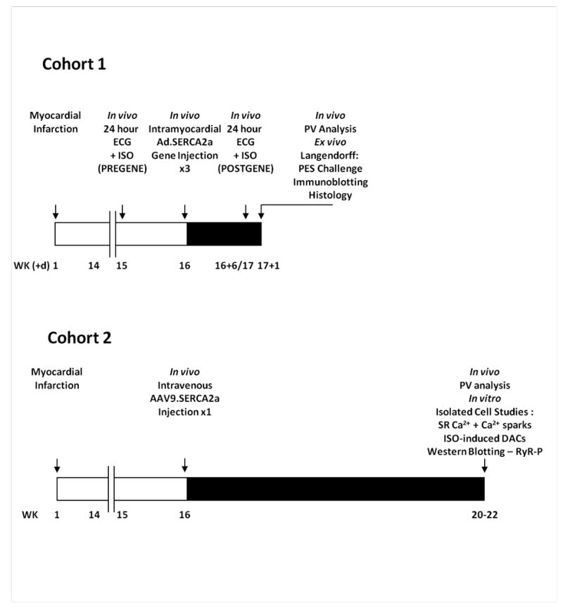
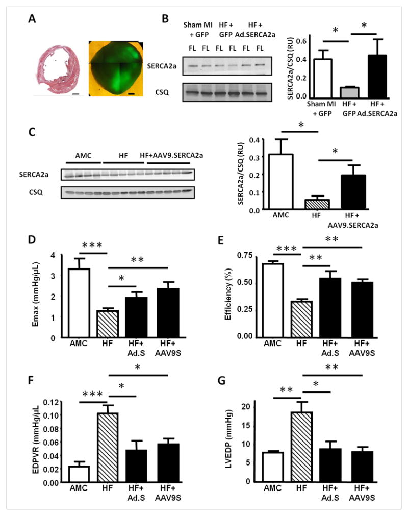
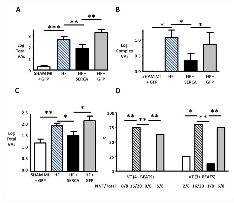
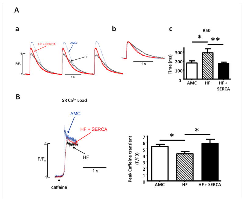
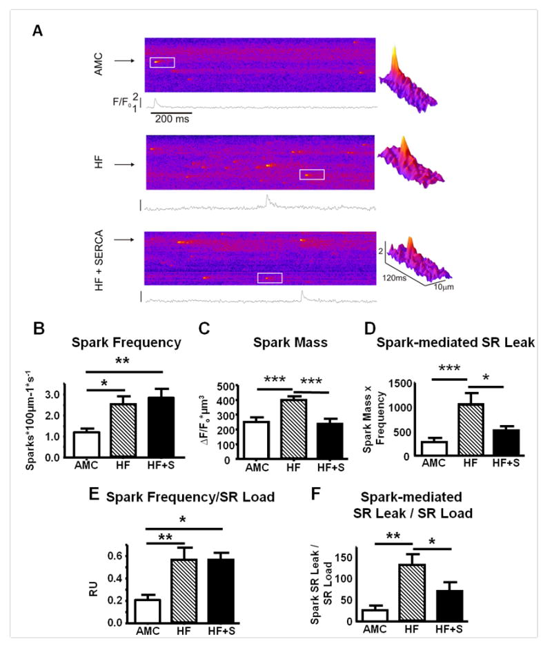
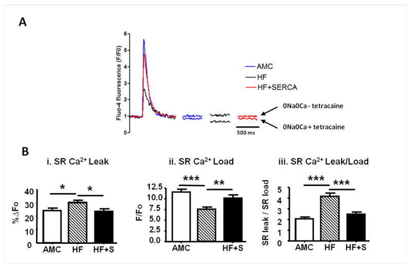
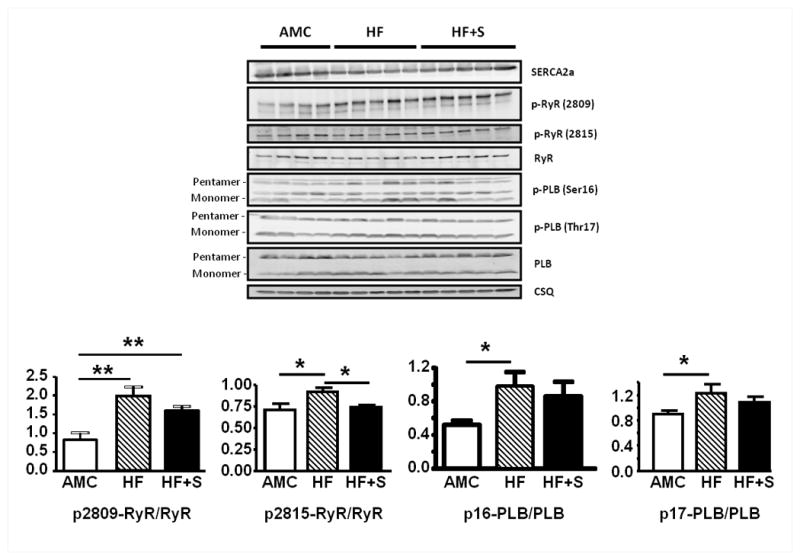
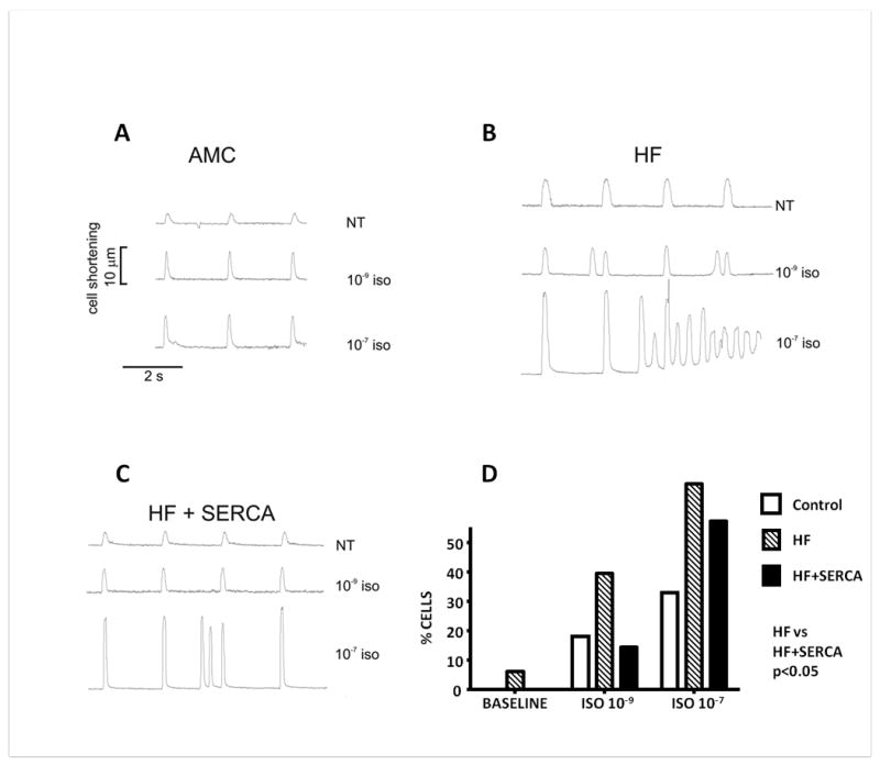
Methods and results: We studied the influence of SERCA2a gene therapy on ventricular arrhythmogenesis in a rat chronic heart failure model. ECG telemetry studies revealed a significant antiarrhythmic effect of SERCA2a gene therapy with reduction of both spontaneous and catecholamine-induced arrhythmias in vivo. SERCA2a gene therapy also reduced susceptibility to reentry arrhythmias in ex vivo programmed electrical stimulation studies. Subcellular Ca(2+) homeostasis and spontaneous SR Ca(2+) leak characteristics were measured in failing cardiomyocytes transfected in vivo with a novel AAV9.SERCA2a vector. SR Ca(2+) leak was reduced after SERCA2a gene therapy, with reversal of the greater spark mass observed in the failing myocytes, despite normalization of SR Ca(2+) load. SERCA2a reduced ryanodine receptor phosphorylation, thereby resetting SR Ca(2+) leak threshold, leading to reduced triggered activity in vitro. Both indirect effects of reverse remodeling and direct SERCA2a effects appear to underlie the antiarrhythmic action.

Conclusions: SERCA2a gene therapy stabilizes SR Ca(2+) load, reduces ryanodine receptor phosphorylation and decreases SR Ca(2+) leak, and reduces cellular triggered activity in vitro and spontaneous and catecholamine-induced ventricular arrhythmias in vivo in failing hearts. SERCA2a gene therapy did not therefore predispose to arrhythmias and may represent a novel antiarrhythmic strategy in heart failure.


Conflict of interest statement

Conflict of Interest Disclosures:RJH is the scientific founder of Celladon Inc which is developing AAV1.SERCA gene therapy for therapeutic purposes.

Figures

Figure 1
Figure 1
Study protocols. Schematic timelines (weeks (+/- days)) summarising the protocols for the two study cohorts evaluating the influence of SERCA2a gene therapy upon arrhythmia generation in a post infarction chronic heart failure model. Cohort 1 – whole heart in vivo and ex vivo studies with Ad.SERCA2a.GFP; Cohort 2 – in vitro cellular and subcellular studies with AAV9.SERCA2a.
Figure 2
Figure 2
Restoration of SERCA2a protein levels after either Ad.SERCA2a or AAV9.SERCA2a gene transfer, with associated improvements in left ventricular function assessed by pressure-volume (PV) analysis in vivo. A. Post infarction heart failure model and SERCA2a gene transfer: (a) Midventricular 10μm section from a chronically infarcted rat heart after staining with haematoxylin and eosin. Scale bar 2mm. (b) A 2mm thick transverse apical section under fluorescence (488nm) microscopy demonstrating GFP expression in two Ad.SERCA2a.GFP injected regions. Scale bar 1mm. B and C. Western blots of SERCA2a and calsequestrin (CSQ) protein in the fluorescent left ventricular myocardium from Ad.SERCA2a gene injected hearts (B), or myocardium from AAV9.SERCA2a injected HF rats (C). D-G. PV measurement of left ventricular Emax (D), energetic efficiency (E), end diastolic PV relationship (EDPVR) (F) and end diastolic pressure (LVEDP) (G). AMC n=6, HF n=8, HF+Ad.SERCA2a (HF+Ad.S) n=5, HF+AAV9SERCA2a (HF+AAV9.S) n=6. *p<0.05, **p<0.01 and ***p<0.001 vs HF.
Figure 3
Figure 3
Reduction of spontaneous and catecholamine-provoked ventricular arrhythmias in failing hearts in vivo after SERCA2a gene transfer. A and B. Spontaneous ventricular arrhythmias during a 24 hour study period. HF rats showed significantly increased spontaneous ventricular arrhythmic events compared with non-failing controls (SHAM MI + GFP). SERCA2a gene transfer significantly reduced the total number of spontaneous ventricular arrhythmias (predominantly ventricular ectopy) (A) and complex ventricular arrhythmias (couplets, triplets and ventricular tachycardia) (B) compared to both untreated HF and HF+GFP cases. C and D. Isoproterenol-induced ventricular arrhythmias during the sixty minute recording period post ISO injection. SERCA2a gene transfer reduced the total number of ISO-induced ventricular arrhythmias (C), and the proportion of cases (%) with ISO-induced ventricular tachycardia (VT) defined as either 4+ or 3+ consecutive ventricular cycles (D). *p<0.05, **p<0.01 and ***p<0.001.
Figure 4
Figure 4
Improvement in cytoplasmic Ca2+ transients, myocyte relaxation kinetics, SR Ca2+ content and basal cytoplasmic Ca2+ levels in failing cardiomyocytes following in vivo AAV9.SERCA2a gene transfer. A. Cytoplasmic Ca2+ transients (AMC (blue), HF (black), HF+SERCA2a (red)) measured from baseline corrected change in emitted fluorescence in fluo4-loaded myocytes stimulated at 0.5Hz (a), with overlain amplitude corrected traces demonstrating difference in Ca2+ transient decay kinetics (b), and quantitative comparison of time (ms) to 50% reduction from peak amplitude (R50) in myocytes paced at 0.5Hz (c). B. SR Ca2+ content in failing myocytes following SERCA2a gene therapy. Representative examples of traces of emitted fluorescence following application of caffeine to determine SR Ca2+ content, and quantitative measurement of SR Ca2+. Number of myocytes (n) studied: AMC n=9, HF n=12, HF+SERCA n=15. *p<0.05 *** p<0.001.
Figure 5
Figure 5
Spontaneous SR Ca2+ release events (sparks) in failing myocytes after SERCA2a gene transfer. A. Representative confocal line scan images demonstrating spontaneous Ca2+ sparks from AMC (upper), HF (middle), and HF+SERCA2a gene transfer (lower) cardiomyocytes. The white inset region is expanded to generate a three dimensional spark representation (shown right), demonstrating the differences in spark morphology after SERCA2a gene transfer. B. Mean spark frequency (number of sparks100μm-1s-1). C. Mean spark mass. D. Spark-mediated SR Ca2+ leak (spark mass × frequency). E-F. Spark frequency (E) and spark-mediated SR Ca2+ leak (F) corrected for matched SR Ca2+ load (mean per heart). Number of sparks/cells/hearts studied: AMC=247/19/7, HF=853/30/11, HF+SERCA=445/10/5. *p<0.05, **p<0.01, ***p<0.001.
Figure 6
Figure 6
SERCA2a gene transfer reduces SR Ca2+ leak and normalises the SR Load-Leak relationship in failing cardiomyocytes. A-B. Measurement of tetracaine-dependent SR Ca2+ leak (using method from Shannon et al). A. Representative examples of Ca2+ transients with paired basal resting levels pre (above) and post (below) 1mM tetracaine (both recorded in 0Na+ 0 Ca2+). B(i). SR Ca2+ leak. B(ii). SR Ca2+ load. B(iii). SR Ca2+ leak per unit load (AMC n=16, HF n=24, HF+S n=13). *p<0.05, ** p<0.01, ***p<0.001.
Figure 7
Figure 7
Protein immunoblotting for SERCA2a, RyR, PLB and CSQ from cohort 2, with levels of PKA- and CaMKII-mediated phosphorylation of RyR and PLB, and quantitative comparison after correction for total RyR and PLB respectively (AMC n=4 HF n=5 HF+S n=5). *p<0.05, ** p<0.01.
Figure 8
Figure 8
Reduction of catecholamine-induced triggered activity in failing cardiomyocytes after SERCA2a gene transfer. A-C. Representative tracings demonstrating contraction profiles of freshly isolated cells field stimulated at 0.5Hz at baseline perfusion with normal Tyrode's solution (NT), and after perfusion with 1 nmol/L and 100 nmol/L Isoproterenol (ISO). A. Age matched controls. B. Failing cardiomyocytes. C. Failing cardiomyocytes four-six weeks post AAV9.SERCA2a gene transfer. D. Proportion of cardiomyocytes from AMC, HF and HF + AAV9.SERCA2a rat hearts demonstrating Ca2+-mediated delayed aftercontractions at baseline, and after treatment with ISO. Number of myocytes studied: AMC=33, HF=33, HF+SERCA=14. Two way ANOVA p<0.05.

References

    1. del Monte F, Harding SE, Schmidt U, Matsui T, Kang ZB, Dec GW, Gwathmey JK, Rosenzweig A, Hajjar RJ. Restoration of contractile function in isolated cardiomyocytes from failing human hearts by gene transfer of SERCA2a. Circulation. 1999;100:2308–11. - PMC - PubMed
    1. del Monte F, Williams E, Lebeche D, Schmidt U, Rosenzweig A, Gwathmey JK, Lewandowski ED, Hajjar RJ. Improvement in survival and cardiac metabolism after gene transfer of sarcoplasmic reticulum Ca(2+)-ATPase in a rat model of heart failure. Circulation. 2001;104:1424–9. - PMC - PubMed
    1. Kawase Y, Ly HQ, Prunier F, Lebeche D, Shi Y, Jin H, Hadri L, Yoneyama R, Hoshino K, Takewa Y, Sakata S, Peluso R, Zsebo K, Gwathmey JK, Tardif JC, Tanguay JF, Hajjar RJ. Reversal of Cardiac Dysfunction After Long-Term Expression of SERCA2a by Gene Transfer in a Pre-Clinical Model of Heart Failure. J Am Coll Cardiol. 2008;51:1112–9. - PubMed
    1. Byrne MJ, Power JM, Preovolos A, Mariani JA, Hajjar RJ, Kaye DM. Recirculating cardiac delivery of AAV2/1SERCA2a improves myocardial function in an experimental model of heart failure in large animals. Gene Ther. 2008;15:1550–7. - PubMed
    1. Hajjar RJ, Zsebo K, Deckelbaum L, Thompson C, Rudy J, Yaroshinsky A, Ly H, Kawase Y, Wagner K, Borow K, Jaski B, London B, Greenberg B, Pauly DF, Patten R, Starling R, Mancini D, Jessup M. Design of a Phase 1/2 Trial of Intracoronary Administration of AAV1/SERCA2a in Patients With Heart Failure. J Cardiac Failure. 2008;14:355–67. - PubMed

Publication types

MeSH terms

Substances