Review
. 2011 Mar 14;16(3):2467-85.
doi: 10.3390/molecules16032467.

Miniproteins as phage display-scaffolds for clinical applications

Affiliations
Review

Miniproteins as phage display-scaffolds for clinical applications

Frederic Zoller et al. Molecules. .

Abstract

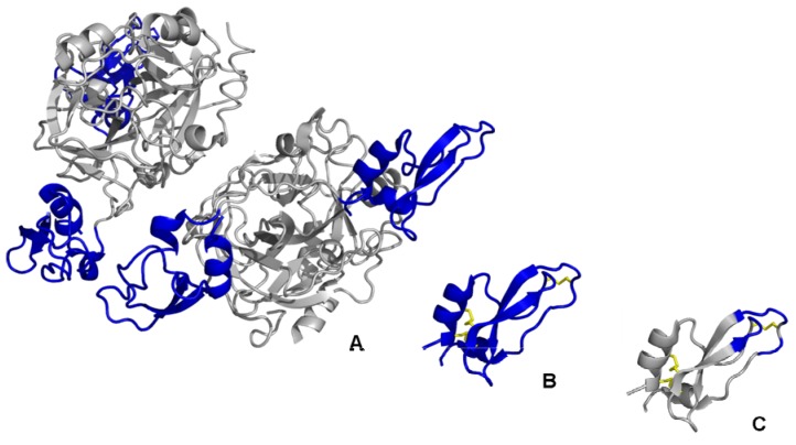
Miniproteins are currently developed as alternative, non-immunoglobin proteins for the generation of novel binding motifs. Miniproteins are rigid scaffolds that are stabilised by alpha-helices, beta-sheets and disulfide-constrained secondary structural elements. They are tolerant to multiple amino acid substitutions, which allow for the integration of a randomised affinity function into the stably folded framework. These properties classify miniprotein scaffolds as promising tools for lead structure generation using phage display technologies. Owing to their high enzymatic resistance and structural stability, miniproteins are ideal templates to display binding epitopes for medical applications in vivo. This review summarises the characteristics and the engineering of miniproteins as a novel class of scaffolds to generate of alternative binding agents using phage display screening. Moreover, recent developments for therapeutic and especially diagnostic applications of miniproteins are reviewed.


Figures

Figure 1
Figure 1
The miniaturisation approach. Downsizing of naturally occurring large protein structures (A), such as the Kunitz-type proteinase inhibitor boophilin (PDB ID: 2ODY), via the isolation of functional protein domains (B) leads to miniature protein scaffolds (C) suitable for engineering for phage display library construction. The functional domains are coloured in blue, disulfide bridges are yellow. Structural representations were created using PyMol software [14].
Figure 2
Figure 2
Schematic representations of different scaffold categories. The first type of scaffold displays a single exposed loop embedded in a ridge template (A). The second scaffold class contains a non-contiguous surface region formed by secondary structural elements (B). The third category shows a complex backbone architecture which encloses multiple discontinuous variable loop segments forming a coherent interface (C). Variable sequence regions are coloured in blue, disulfide bridges are coloured in yellow. The schematic representations were generated from the RCSB Protein Data Base: (A): Scorpion charybdotoxin (PDB ID: 2CRD); (B): Single zinc finger DNA-binding domain (PDB ID: 1ZNF); (C): Cellulose binding domain from cellobiohydrolase CeI7A (PDB ID: 1CBH). Structural representations were illustrated using PyMol software [14].
Figure 3
Figure 3
Schematic representation of Ecballium elaterium trypsin inhibitor II (left) and its functional miniature scaffold Min-23 (right), showing the cystine-stabilised β-sheet (CSB) motif of the knottin family (disulfide bridges are coloured in yellow). The single exposed variable loop (coloured in blue) is permissive to the variation of six amino acids in EETI-II and 8 to 10 in Min-23, respectively. Structural representations (PDB ID: 2IT7) were illustrated using PyMol software [14].
Figure 4
Figure 4
Schematic representation of the Z domain scaffold, termed Affibody, derived from the B-domain of the Staphylococcal aureus immunoglobulin-binding protein A. The composed surface formed by the three-α-helical structure (coloured in blue) presents 13 variable amino acid positions in two α-helices for combinatorial randomisation via phage display. The structural data (PDB ID: 2B89) were illustrated using PyMol software [14].
Figure 5
Figure 5
Schematic representations of tendamistat (A) and the Kunitz-domain scaffold (B). The discontinuous surface domains form a coherent interface for the target interaction (coloured in blue, disulfide bridges are coloured in yellow). The structural data (PDB ID: (A): 1OK0; (B): 1AAP) were illustrated using PyMol software [14].

Similar articles

Cited by

References

    1. Skerra A. Imitating the humoral immune response. Curr. Opin. Chem. Biol. 2003;7:683–693. doi: 10.1016/j.cbpa.2003.10.012. - DOI - PubMed
    1. Binz H.K., Amstutz P., Pluckthun A. Engineering novel binding proteins from nonimmunoglobulin domains. Nat. Biotechnol. 2005;23:1257–1268. doi: 10.1038/nbt1127. - DOI - PubMed
    1. Descotes J. Immunotoxicity of monoclonal antibodies. mAbs. 2009;1:104–111. doi: 10.4161/mabs.1.2.7909. - DOI - PMC - PubMed
    1. Wu A.M., Senter P.D. Arming antibodies: Prospects and challenges for immunoconjugates. Nat. Biotechnol. 2005;23:1137–1146. doi: 10.1038/nbt1141. - DOI - PubMed
    1. Holliger P., Hudson P.J. Engineered antibody fragments and the rise of single domains. Nat. Biotechnol. 2005;23:1126–1136. - PubMed

Publication types

LinkOut - more resources