. 2011 Aug;19(8):875-81.
doi: 10.1038/ejhg.2011.42. Epub 2011 Mar 16.

Novel mutations affecting LRP5 splicing in patients with osteoporosis-pseudoglioma syndrome (OPPG)

Affiliations

Novel mutations affecting LRP5 splicing in patients with osteoporosis-pseudoglioma syndrome (OPPG)

C M Laine et al. Eur J Hum Genet. 2011 Aug.

Abstract

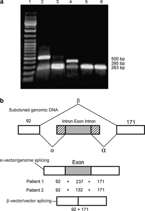
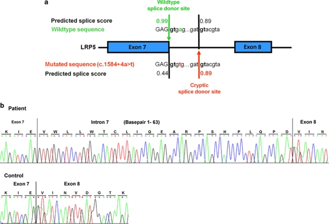
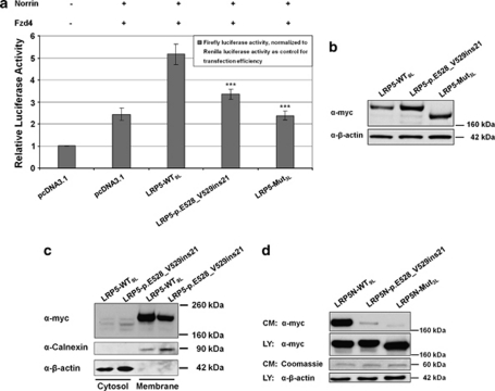
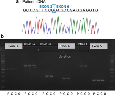
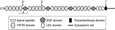
Osteoporosis-pseudoglioma sydrome (OPPG) is an autosomal recessive disorder with early-onset severe osteoporosis and blindness, caused by biallelic loss-of-function mutations in the low-density lipoprotein receptor-related protein 5 (LRP5) gene. Heterozygous carriers exhibit a milder bone phenotype. Only a few splice mutations in LRP5 have been published. We present clinical and genetic data for four patients with novel LRP5 mutations, three of which affect splicing. Patients were evaluated clinically and by radiography and bone densitometry. Genetic screening of LRP5 was performed on the basis of the clinical diagnosis of OPPG. Splice aberrances were confirmed by cDNA sequencing or exon trapping. The effect of one splice mutation on LRP5 protein function was studied. A novel splice-site mutation c.1584+4A>T abolished the donor splice site of exon 7 and activated a cryptic splice site, which led to an in-frame insertion of 21 amino acids (p.E528_V529ins21). Functional studies revealed severely impaired signal transduction presumably caused by defective intracellular transport of the mutated receptor. Exon trapping was used on two samples to confirm that splice-site mutations c.4112-2A>G and c.1015+1G>T caused splicing-out of exons 20 and 5, respectively. One patient carried a homozygous deletion of exon 4 causing the loss of exons 4 and 5, as demonstrated by cDNA analysis. Our results broaden the spectrum of mutations in LRP5 and provide the first functional data on splice aberrations.


Figures

Figure 1
Figure 1
Splicing assay with the exon trapping assay. (a) Electrophoresis of cDNA-PCR products generated from the wild-type and mutant constructs (exon 20 from patient 1 and exon 5 from patient 2) after transfection into COS-7 cells. Lane 1: DNA marker, GeneRuler DNA Ladder Mix (MBI Fermentas, Glen Burnie, MD, USA); lane 2: splicing product of exon 20 wild-type construct; lane 3: splicing product of exon 20 mutant construct; lane 4: splicing product of exon 5 wild-type construct; lane 5: splicing product of exon 5 mutant construct; lane 6: splicing product of pSPL3. In the absence of an inserted fragment, a vector/vector splice product of 263 bp is produced (A: lane 6, B: β splice product). In the presence of an inserted wild-type fragment, splicing of the vector gives rise to two products: a vector/vector product of 263 bp and a correctly spliced vector/genomic product containing the inserted exon (237 or 132 bp; A: lanes 2 and 4, B: α). The abundance of vector/genome product as compared with the vector/vector product indicates that no enhancement of exon 20- or 5-skipping occurred in the wild-type constructs. Mutant constructs did not produce any detectable vector/genomic product from the mutant inserts obtained from patients 1 and 2 (lanes 3 and 5), indicating complete splicing-out of exons 20 or 5. (b) Genomic DNA constructs showing the cloned sequence within the exon-trapping vector, pSPL3. The sizes of vector DNA (white box), intronic DNA (lines) and exonic DNA (black box with white dots) are indicated. Two possible splicing pathways, α (vector/genome) and β (vector/vector) are shown with their respective spliced products.
Figure 2
Figure 2
Identification of the c.1584+4A>T disease-causing splice-site mutation in LRP5. (a) Direct-sequencing results of genomic DNA amplicons of the LRP5 exon 7 splice donor site in a control individual (wild-type sequence) and patient 3A (mutated sequence). The predicted splice score (0.99) of the wild-type splice donor site decreases to 0.44 in the mutated sequence, inducing the activation of a cryptic splice donor site downstream. (b) Electropherograms of LRP5 cDNA amplicons from patient 3A (upper panel) and a control individual (lower panel) revealing a mutated splice donor site after exon 7, with 63 additional nucleotides of intron 7 inserted into the RNA molecule in the patient's sample. The wild-type sequence is correctly spliced in the control individual.
Figure 3
Figure 3
Functional studies with mutant LRP5 protein. (a) Dual-luciferase reporter assay measuring the canonical Wnt signaling activity. The co-transfected Firefly luciferase reportergen construct (Topflash TCF reporter plasmid) contains TCF-binding sites as a target for activated canonical Wnt signaling. The graph describes the relative luciferase activities (average and standard deviation) of empty vector (pcDNA3.1), wild-type LRP5 (LRP5-WT9L), and the mutant LRP5-p.E528_V529ins21. The mutant LRP5-Mut3L serves as a positive control for impaired Wnt/Norrin signal transduction. Note that activities of the mutant LRP5-p.E528_V529ins21 are significantly lower than those of LRP5-WT9 L (P-values <0.001 (***) in a two-sided Student's t-test calculation). (b) Western blot of cell lysates from transiently transfected HEK293T cells used in the dual-luciferase reporter assay (results presented in a), confirming expression of WT9L and of the two mutated LRP5 receptors. (c) Cell fractioning assay. HEK293T cells were transiently transfected with LRP5-WT9L or LRP5-p.E528_V529ins21. Both WT9L and p.E528_V529ins21 are almost exclusively detectable in the membrane fraction. (d) Secretion assay. Representative western blot of conditioned medium (CM) and cell lysate (LY) from HEK293T cells transiently transfected with LRP5N-WT9L, LRP5N-p.E528_V529ins21 and LRP5N-Mut3L expression constructs. Comparable loading of cell lysate or conditioned medium across experiments is indicated by immunodetection of β-actin or by Coomassie staining of a 60-kDa protein in the conditioned medium, respectively. Note that only LRP5N-WT9L is strongly detected in conditioned medium.
Figure 4
Figure 4
(a) Electropherograms of LRP5 cDNA amplicons from patient 4 revealed the loss of exons 4 and 5. (b) PCR-amplified fragments from genomic DNA of patient 4 and two controls visualized on a 2% ethidium bromide gel. All studied amplicons; exons 3, 4 and 5, areas of intron 3 (a: 2735–3671 nucleotides and b: 4198–4503 nucleotides downstream of exon 3) and intron 4 (607–1213 nucleotides downstream of exon 4) can be detected in both control samples (C) and are clean in water samples (0) serving as negative controls. PCR of the patient's (P) genomic DNA was repeatedly unsuccessful in the areas of intron 3b, exon 4 and intron 4, suggesting a homozygous deletion involving exon 4 and adjacent intronic parts. Exon 5 and at least 10 nucleotides of the adjoining introns could be sequenced from both patient and control samples.
Figure 5
Figure 5
Schematic representation of the LRP5 protein and the areas affected by the mutations presented in this study. (1) The splicing-out of exon 20 in patient 1 abolishes the entire transmembrane domain of the protein. (2) The in-frame splicing-out of exon 5 in patient 2 induces a stop codon at the beginning of exon 5, which encodes the first EGF repeat. (3) The insertion of 21 amino acids after exon 7 in patient 3 affects the second β-propeller between two YTWD domains. (4) The loss of exons 4 and 5 in patient 4 causes the loss of the last YWTD domain in the first β-propeller and the entire first EGF repeat.

References

    1. Ai M, Heeger S, Bartels CF, Schelling DK. Clinical and molecular findings in osteoporosis-pseudoglioma syndrome. Am J Hum Genet. 2005;77:741–753. - PMC - PubMed
    1. Gong Y, Vikkula M, Boon L, et al. Osteoporosis-pseudoglioma syndrome, a disorder affecting skeletal strength and vision, is assigned to chromosome region 11q12–13. Am J Hum Genet. 1996;59:146–151. - PMC - PubMed
    1. Somer H, Palotie A, Somer M, Hoikka V, Peltonen L. Osteoporosis-pseudoglioma syndrome: clinical, morphological, and biochemical studies. J Med Genet. 1988;25:543–549. - PMC - PubMed
    1. Beighton P. Osteoporosis-pseudoglioma syndrome. Clin Genet. 1986;29:263. - PubMed
    1. Frontali M, Dallapiccola B. Osteoporosis-pseudoglioma syndrome and the ocular form of osteogenesis imperfecta. Clin Genet. 1986;29:262. - PubMed

Publication types

Substances

Supplementary concepts