. 2011 Feb 23;6(2):e17046.
doi: 10.1371/journal.pone.0017046.

From attachment to damage: defined genes of Candida albicans mediate adhesion, invasion and damage during interaction with oral epithelial cells

Affiliations

From attachment to damage: defined genes of Candida albicans mediate adhesion, invasion and damage during interaction with oral epithelial cells

Betty Wächtler et al. PLoS One. .

Abstract

Candida albicans frequently causes superficial infections by invading and damaging epithelial cells, but may also cause systemic infections by penetrating through epithelial barriers. C. albicans is an unusual pathogen because it can invade epithelial cells via two distinct mechanisms: induced endocytosis, analogous to facultative intracellular enteropathogenic bacteria, and active penetration, similar to plant pathogenic fungi. Here we investigated the molecular basis of C. albicans epithelial interactions. By systematically assessing the contributions of defined fungal pathways and factors to different stages of epithelial interactions, we provide an expansive portrait of the processes and activities involved in epithelial infection. We strengthen the concept that hyphal formation is critical for epithelial invasion. Importantly, our data support a model whereby initial epithelial invasion per se does not elicit host damage, but that C. albicans relies on a combination of contact-sensing, directed hyphal extension, active penetration and the expression of novel pathogenicity factors for further inter-epithelial invasion, dissemination and ultimate damage of host cells. Finally, we explore the transcriptional landscape of C. albicans during the early stages of epithelial interaction, and, via genetic analysis, identify ICL1 and PGA34 as novel oral epithelial pathogenicity factors.


Conflict of interest statement

Competing Interests: The authors have declared that no competing interests exist.

Figures

Figure 1
Figure 1. Groups of mutants lacking genes associated with adhesion, invasion and damage.
Adhesion, invasion and damage potential was determined for each mutant and normalized against the relevant wild type. Group 2a mutants showed reduced adhesion, invasion and damage. The group 2b mutants efg1Δ and ecm33Δ show significant reduction in adhesion, but have a further decreased ability to invade into epithelial cells, suggesting distinct adhesion and invasion functions of these genes. The group 3 mutant pmt2Δ/PMT2 lacks factors or activities which are specifically associated with invasion and damage, but not adhesion. Group 4 mutants lacking MKC1, GPP1, GPD2 and EED1 showed significant reduction in damage, but not adhesion and invasion. Group 5 mutants lacking CKA2, BCR1, HWP1, BUD2 or RSR1 showed significant reduction in adhesion and damage, but not invasion. n. d.  =  not determined; */**, significant difference compared to the corresponding wild type (WT) (p<0.05/p<0.01). Note that adhesion, invasion and damage assays were performed separately.
Figure 2
Figure 2. Adhesion kinetics of C. albicans wild type (WT), bud2Δ, rsr1Δ and hwp1Δ mutant cells.
Oral TR146 epithelial cells were co-incubated with 105 C. albicans cells for either 20, 60, 120, or 180 min. After extensive washing and fixation, the samples were stained and the adherent cells were counted under the fluorescence microscope. The experiment was performed at least three times in duplicates. The values are calculated as percentage of adherent cells compared to wild type adherent cells at 180 min (100%). */**, significant difference compared to the adhesion of the corresponding WT to oral epithelial cells (p<0.05/p<0.01).
Figure 3
Figure 3. Enterocyte invasion by group 4 and 5 damage-associated mutants.
C. albicans cells (wild type and rsr1Δ, bud2Δ, hwp1Δ, bcr1Δ, cka2Δ, eed1Δ, gpp1Δ, gpd2Δ and mkc1Δ) were co-incubated with Caco-2 enterocytes for 3 h. After fixation the samples were differentially stained and analyzed under the fluorescence microscope. The experiment was performed at least three times in duplicates. */**, significant difference compared to the corresponding WT invasion (control) (p<0.05/p<0.01).
Figure 4
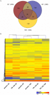
Figure 4. Distribution of differentially regulated genes and hierarchical clustering.
(A) Venn diagram showing the distribution of the differentially regulated genes from C. albicans cells grown on epithelial monolayer at 20, 60 and 180 min after horizontal analysis as compared to plastic. (B) Hierarchical clustering of C. albicans genes expressed at 20, 60 and 180 min incubation on oral epithelial cells versus plastic. Gene expression is shown in a logarithmic color range from −11.31 in blue to 11.31 in red. Non regulated genes (1) are shown in yellow. The profiles of the earliest time points cluster more distant to the later time points. The expression profiles of C. albicans exposed to epithelial cells at 20, 60 and 180 min did not show any clear similarities. In contrast, the profiles of cells exposed to plastic for 60 and 180 min were most similar to each other as compared to all other samples.
Figure 5
Figure 5. Adhesion, invasion and damage properties of selected mutants lacking epithelial infection up-regulated genes.
C. albicans mutant strains lacking ICL1 and PGA34 showed wild type levels of adhesion and/or invasion. However, icl1Δ showed significantly reduced invasion and damage and pga34Δ showed significantly reduced damage. The values are calculated as percentage of adherent/invasive cells or damage compared to the corresponding wild type (100%). **, significant difference compared to the corresponding wild type (WT) (p<0.01).

References

    1. Mavor AL, Thewes S, Hube B. Systemic fungal infections caused by Candida species: epidemiology, infection process and virulence attributes. Curr Drug Targets. 2005;6:863–874. - PubMed
    1. Odds FC. Baltimore: University Park Press; 1988. Ecology and epidemiology of candidiasis.
    1. Ruhnke M. Washington DC: American Society for Microbiology Press; 2002. Skin and mucous membrane infections. pp. 307–325.
    1. Calderone R. Washington, DC: ASM Press; 2002. Candida and Candidiasis.
    1. Hube B. From commensal to pathogen: stage- and tissue-specific gene expression of Candida albicans. Curr Opin Microbiol. 2004;7:336–341. - PubMed

Publication types